Application
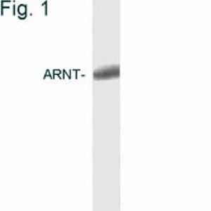
Western blot of HIF1 beta in COS cell extract.Application
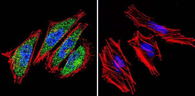
Immunofluorescent analysis of HIF1 beta in A2058 cells. HIF1 beta (green), F-Actin staining with Phalloidin (red) and nuclei with DAPI (blue) is shown. Cells were grown on slides and fixed with formaldehyde prior to staining. Cells were probed without (control) or with HIF1 beta antibody [2B10] at a dilution of 1:100 over night at 4 °C, washed with PBS and incubated with a proper secondary antibody. Images were taken at 60X magnification.| Product Name | HIF1 beta antibody |
|---|---|
| Antibody Type | Primary Antibodies |
| Product description | Mouse monoclonal antibody [2B10] to HIF1 betaThe aryl hydrocarbon (Ah) receptor is involved in the induction of several enzymes that participate in xenobiotic metabolism. The ligand-free, cytosolic form of the Ah receptor is complexed to heat shock protein 90. Binding of ligand, which includes dioxin and polycyclic aromatic hydrocarbons, results in translocation of the ligand-binding subunit only to the nucleus. Induction of enzymes involved in xenobiotic metabolism occurs through binding of the ligand-bound Ah receptor to xenobiotic responsive elements in the promoters of genes for these enzymes. This gene encodes a protein that forms a complex with the ligand-bound Ah receptor, and is required for receptor function. The encoded protein has also been identified as the beta subunit of a heterodimeric transcription factor, hypoxia-inducible factor 1. A t(1;12)(q21;p13) translocation, which results in a TEL-ARNT fusion protein, is associated with acute myeloblastic leukemia. Alternative splicing results in multiple transcript varia |
| Immunogen | Synthetic peptide corresponding to residues C N(771) S Y N N E E F P D L T M F P P F S E(789) of human ARNT. |
| Clonality | Monoclonal |
|---|---|
| Isotype | IgG1 |
| Clone Number | 2B10 |
| Host Species | Mouse |
| Tested Applications | GSAICC/IFIHCIHC-PIPWB |
| IHC-P: 5 µg/ml: |
|
| Species Reactivity | HumanMouseRatZebra FishFishPrimateXenopus |
| Concentration | 1mg/ml (Please refer to the vial label for the specific concentration) |
| Gene Synonyms | HIF-1-beta ARNT HIF1-beta bHLHe2 aryl hydrocarbon receptor nuclear translocator HIF-1beta HIF1BETA TANGO HIF1B |
|---|---|
| Target | HIF-1 beta |
Application
Western blot of HIF1 beta in COS cell extract.
Application
Immunofluorescent analysis of HIF1 beta in A2058 cells. HIF1 beta (green), F-Actin staining with Phalloidin (red) and nuclei with DAPI (blue) is shown. Cells were grown on slides and fixed with formaldehyde prior to staining. Cells were probed without (control) or with HIF1 beta antibody [2B10] at a dilution of 1:100 over night at 4 °C, washed with PBS and incubated with a proper secondary antibody. Images were taken at 60X magnification.| Application Notes | IHC-P: 5 µg/ml: |
|---|
| Form | Liquid |
|---|---|
| Storage Instructions | -20° C, Avoid Freeze/Thaw Cycles |
| Storage Buffer | PBS containing 0.05 % sodium azide and 1 mg/ml BSA |


