Application
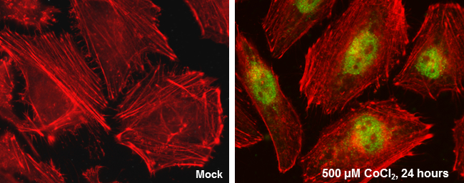
HIF2 alpha antibody [GT125] detects HIF2 alpha protein at nucleus by immunofluorescent analysis. Sample: HeLa cells were fixed in 4% paraformaldehyde at RT for 15 min. Green: HIF2 alpha protein stained by HIF2 alpha antibody [GT125] diluted at 1:100. Red: Actin filaments were labeled with Phalloidin.Application
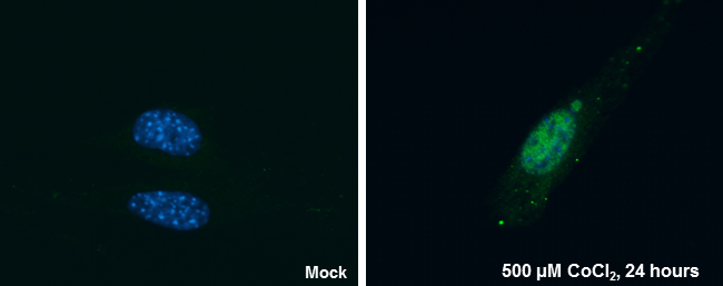
HIF2 alpha antibody [GT125] detects HIF2 alpha protein at nucleus by immunofluorescent analysis. Sample: NIH/3T3 cells were fixed in 4% paraformaldehyde at RT for 15 min. Green: HIF2 alpha protein stained by HIF2 alpha antibody [GT125] diluted at 1:100. Blue: Hoechst 33342 staining.Application
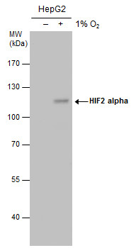
HIF2 alpha antibody detects HIF2 alpha protein by western blot analysis. Un-treated (-) and treated (+, 1% O2 treatment for 24hr) HepG2 whole cell extracts (30 µg) were separated by 7.5% SDS-PAGE, and the membrane was blotted with HIF2 alpha antibody at a dilution of 1:500. The HRP-conjugated anti-mouse IgG antibody was used to detect the primary antibody.Application
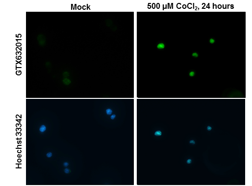
HIF2 alpha antibody [GT125] detects HIF2 alpha protein at nucleus by immunofluorescent analysis. Samples: PC-12 cells mock (left) and treated with 500 μM CoCl2 for 24 hrs (right) were fixed in 4% paraformaldehyde at RT for 15 min. Green: HIF2 alpha protein stained by HIF2 alpha antibody [GT125] diluted at 1:100. Blue: Hoechst 33342 staining.Application
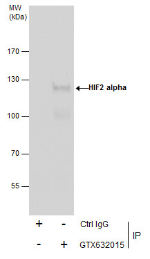
Immunoprecipitation of HIF2 alpha protein from HepG2 whole cell extracts treated with 500 μM CoCl2 for 24 hr using 5 μg of HIF2 alpha antibody [GT125] . Western blot analysis was performed using HIF2 alpha antibody [GT125] . EasyBlot anti-Mouse IgG was used as a secondary reagent.| Product Name | HIF2 alpha antibody |
|---|---|
| Antibody Type | Primary Antibodies |
| Product description | Mouse Monoclonal antibody [GT125] to HIF2 alpha (endothelial PAS domain protein 1)This gene encodes a transcription factor involved in the induction of genes regulated by oxygen, which is induced as oxygen levels fall. The encoded protein contains a basic-helix-loop-helix domain protein dimerization domain as well as a domain found in proteins in signal transduction pathways which respond to oxygen levels. Mutations in this gene are associated with erythrocytosis familial type 4. [provided by RefSeq] |
| Immunogen | Recombinant protein encompassing a sequence within the center region of human HIF2 alpha. The exact sequence is proprietary. |
| Clonality | Monoclonal |
|---|---|
| Isotype | IgG1 |
| Clone Number | GT125 |
| Host Species | Mouse |
| Tested Applications | ICC/IFIPWB |
| *Optimal dilutions/concentrations should be determined by the researcher.
: |
|
| Species Reactivity | HumanMouseRat |
| Predicted Reactivity | PigPig (83%) |
| Concentration | 1mg/ml |
| Purification | Affinity purified |
| Gene Synonyms | ECYT4 PASD2 bHLHe73 endothelial PAS domain protein 1 HIF2A HLF EPAS1 MOP2 |
|---|---|
| Target | HIF2 alpha |
| Molecular Weight(MW) | 96 kDa (note) |
| Cellular Localization | Nucleus |
Application
HIF2 alpha antibody [GT125] detects HIF2 alpha protein at nucleus by immunofluorescent analysis. Sample: HeLa cells were fixed in 4% paraformaldehyde at RT for 15 min. Green: HIF2 alpha protein stained by HIF2 alpha antibody [GT125] diluted at 1:100. Red: Actin filaments were labeled with Phalloidin.
Application
HIF2 alpha antibody [GT125] detects HIF2 alpha protein at nucleus by immunofluorescent analysis. Sample: NIH/3T3 cells were fixed in 4% paraformaldehyde at RT for 15 min. Green: HIF2 alpha protein stained by HIF2 alpha antibody [GT125] diluted at 1:100. Blue: Hoechst 33342 staining.
Application
HIF2 alpha antibody detects HIF2 alpha protein by western blot analysis. Un-treated (-) and treated (+, 1% O2 treatment for 24hr) HepG2 whole cell extracts (30 µg) were separated by 7.5% SDS-PAGE, and the membrane was blotted with HIF2 alpha antibody at a dilution of 1:500. The HRP-conjugated anti-mouse IgG antibody was used to detect the primary antibody.
Application
HIF2 alpha antibody [GT125] detects HIF2 alpha protein at nucleus by immunofluorescent analysis. Samples: PC-12 cells mock (left) and treated with 500 μM CoCl2 for 24 hrs (right) were fixed in 4% paraformaldehyde at RT for 15 min. Green: HIF2 alpha protein stained by HIF2 alpha antibody [GT125] diluted at 1:100. Blue: Hoechst 33342 staining.
Application
Immunoprecipitation of HIF2 alpha protein from HepG2 whole cell extracts treated with 500 μM CoCl2 for 24 hr using 5 μg of HIF2 alpha antibody [GT125] . Western blot analysis was performed using HIF2 alpha antibody [GT125] . EasyBlot anti-Mouse IgG was used as a secondary reagent.| Positive Control | HepG2 (1% O2 treatment for 24hr) |
|---|---|
| Application Notes | *Optimal dilutions/concentrations should be determined by the researcher.
: |
| Form | Liquid |
|---|---|
| Storage Instructions | Keep as concentrated solution. Aliquot and store at -20ºC or below. Avoid multiple freeze-thaw cycles. |
| Storage Buffer | PBS |


