Application
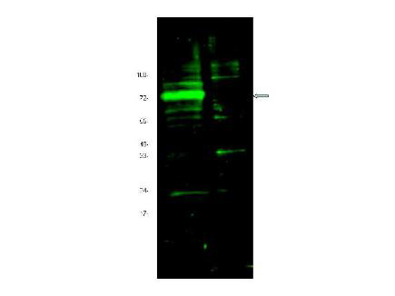
Anti-Hif Antibody - Western Blot. Western blot of Affinity Purified anti-Hif antibody shows detection of a band ~72 kD corresponding to mouse Hif3a (arrowhead). Approximately 10 ug of a CoCl2 treated 3T3 cell lysate (lane 1) and control 3T3 cell lysate (lane 2) were separated by 4-20% SDS-PAGE and transferred onto nitrocellulose. Treatment of exponentially growing 3T3 cells with 130 uM CoCl2 for 6 h at 37? C effectively mimics hypoxia. After blocking the membrane was probed overnight at 4C with the primary antibody diluted to 1:1600. The membrane was washed and reacted with a 1:10000 dilution of IRDye800 conjugated Gt-a-Rabbit IgG [H&L] MX ( for 45 min at room temperature. IRDye800 fluorescence image was captured using the Odyssey Infrared Imaging System developed by LI-COR. IRDye is a trademark of LI-COR, Inc. Other detection systems will yield similar results.Application
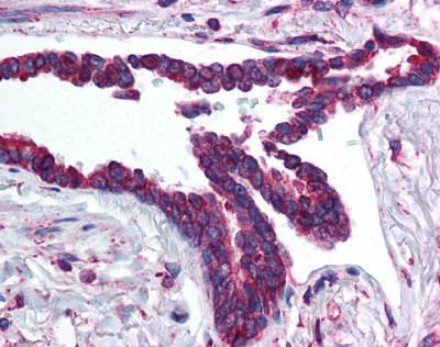
Human Lung, Respiratory Epithelium (formalin-fixed, paraffin-embedded) stained with HIF3A antibody at 5 ug/ml followed by biotinylated goat anti-rabbit IgG secondary antibody LS-D1, alkaline phosphatase-streptavidin and chromogen.Application
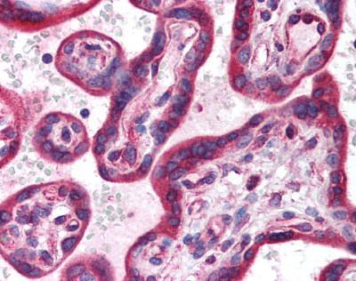
Human Placenta (formalin-fixed, paraffin-embedded) stained with HIF3A antibody at 5 ug/ml followed by biotinylated goat anti-rabbit IgG secondary antibody LS-D1, alkaline phosphatase-streptavidin and chromogen.| Product Name | HIF3 alpha antibody |
|---|---|
| Antibody Type | Primary Antibodies |
| Product description | Rabbit polyclonal antibody to HIF3A |
| Immunogen | Synthetic peptide - KLH conjugated |
| Clonality | Polyclonal |
|---|---|
| Host Species | Rabbit |
| Tested Applications | ELISAICC/IFIHC-PWB |
| WB (1:100 - 1:8000), ELISA (1:14000 - 1:80000), IF, IHC-P (5 µg/ml): |
|
| Species Reactivity | HumanMouseRat |
| Concentration | 1mg/ml (Please refer to the vial label for the specific concentration) |
| Purification | Affinity purified |
| Gene Synonyms | Ipas MOP7 NEPAS bHLHe17 |
|---|---|
| Tissue Specificity | aa 581-592 of mouse Hif3 a (Hypoxia Inducible Factor). |
Application
Anti-Hif Antibody - Western Blot. Western blot of Affinity Purified anti-Hif antibody shows detection of a band ~72 kD corresponding to mouse Hif3a (arrowhead). Approximately 10 ug of a CoCl2 treated 3T3 cell lysate (lane 1) and control 3T3 cell lysate (lane 2) were separated by 4-20% SDS-PAGE and transferred onto nitrocellulose. Treatment of exponentially growing 3T3 cells with 130 uM CoCl2 for 6 h at 37? C effectively mimics hypoxia. After blocking the membrane was probed overnight at 4C with the primary antibody diluted to 1:1600. The membrane was washed and reacted with a 1:10000 dilution of IRDye800 conjugated Gt-a-Rabbit IgG [H&L] MX ( for 45 min at room temperature. IRDye800 fluorescence image was captured using the Odyssey Infrared Imaging System developed by LI-COR. IRDye is a trademark of LI-COR, Inc. Other detection systems will yield similar results.
Application
Human Lung, Respiratory Epithelium (formalin-fixed, paraffin-embedded) stained with HIF3A antibody at 5 ug/ml followed by biotinylated goat anti-rabbit IgG secondary antibody LS-D1, alkaline phosphatase-streptavidin and chromogen.
Application
Human Placenta (formalin-fixed, paraffin-embedded) stained with HIF3A antibody at 5 ug/ml followed by biotinylated goat anti-rabbit IgG secondary antibody LS-D1, alkaline phosphatase-streptavidin and chromogen.| Application Notes | WB (1:100 - 1:8000), ELISA (1:14000 - 1:80000), IF, IHC-P (5 µg/ml): |
|---|
| Form | Liquid |
|---|---|
| Storage Instructions | +4°C or -20°C, Avoid repeated freezing and thawing. |
| Storage Buffer | 0.02 M potassium phosphate, 0.15 M sodium chloride, pH 7.2, 0.01% sodium azide. |


