Application
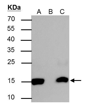
Histone H2BK20ac (acetyl Lys20) antibody immunoprecipitates Histone H2BK20ac (acetyl Lys20) protein in IP experiments. IP Sample: HeLa whole cell lysate/extract A : 30 μg whole cell lysate/extract of Histone H2BK20ac (acetyl Lys20) protein expressing HeLa cells (0.4uM TSA treatment for 18hr) B : Control with 2.5 μg of pre-immune rabbit IgG C : Immunoprecipitation of Histone H2BK20ac (acetyl Lys20) protein by 2.5 μg of Histone H2BK20ac (acetyl Lys20) antibody 12% SDS-PAGE The immunoprecipitated Histone H2BK20ac (acetyl Lys20) protein was detected by Histone H2BK20ac (acetyl Lys20) antibody diluted at 1 : 5000. EasyBlot anti-rabbit IgG (HRP) was used as a secondary reagent.Application
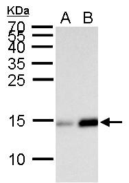
Histone H2BK20ac (acetyl Lys20) antibody detects Histone H2BK20ac (acetyl Lys20) protein by Western blot analysis. A. 30 μg HeLa whole cell lysate/extract (0.012% DMSO treatment for 18 hr) B. 30 μg HeLa whole cell lysate/extract (0.4 μM Trichostatin A treatment for 18 hr) 15 % SDS-PAGE Histone H2BK20ac (acetyl Lys20) antibody dilution: 1:5000Application
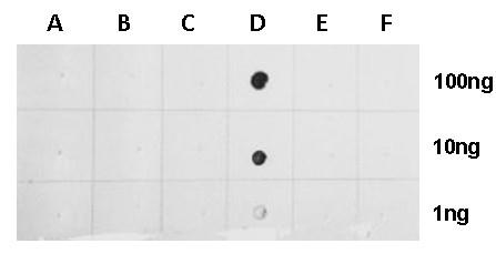
Dotblot analysis of anti-Histone H2BK20ac (acetyl Lys20) antibody with peptide samples. Varied amount of peptide samples were spotted onto positively charged nylon membrane and blotted with Histone H2BK20ac (acetyl Lys20) antibody 1:5000 dilution. A: Peptide samples of H2A.2K5 un B: Peptide samples of H2A.2K5ac C: Peptide samples of H2BK20 un D: Peptide samples of H2BK20ac E: Peptide samples of H2BK5 un F: Peptide samples of H2BK5acApplication
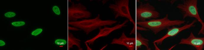
Histone H2BK20ac (acetyl Lys20) antibody detects Histone H2BK20ac (acetyl Lys20) protein at nucleus by immunofluorescent analysis. Sample: 0.4M TSA-treated for 18hr HeLa cells were fixed in 4% paraformaldehyde at RT for 15 min. Green: Histone H2BK20ac (acetyl Lys20) protein stained by Histone H2BK20ac (acetyl Lys20) antibody diluted at 1:500. Red: alpha Tubulin antibody [GT114], a cytoskeleton marker, stained by diluted at 1:500. Blue: Hoechst 33342 staining.| Product Name | Histone H2BK20ac (acetyl Lys20) antibody |
|---|---|
| Antibody Type | Primary Antibodies |
| Product description | Rabbit Polyclonal antibody to Histone H2B (acetyl Lys20) |
| Modification | acetyl Lys20 |
| Clonality | Polyclonal |
|---|---|
| Isotype | IgG |
| Host Species | Rabbit |
| Tested Applications | Dot,ICC/IF,IP,WB |
| *Optimal dilutions/concentrations should be determined by the researcher. | |
| Species Reactivity | Human |
| Concentration | 1mg/ml (Please refer to the vial label for the specific concentration) |
| Purification | Affinity purified |
| Target | Histone H2B (acetyl Lys20) |
|---|
Application
Histone H2BK20ac (acetyl Lys20) antibody immunoprecipitates Histone H2BK20ac (acetyl Lys20) protein in IP experiments. IP Sample: HeLa whole cell lysate/extract A : 30 μg whole cell lysate/extract of Histone H2BK20ac (acetyl Lys20) protein expressing HeLa cells (0.4uM TSA treatment for 18hr) B : Control with 2.5 μg of pre-immune rabbit IgG C : Immunoprecipitation of Histone H2BK20ac (acetyl Lys20) protein by 2.5 μg of Histone H2BK20ac (acetyl Lys20) antibody 12% SDS-PAGE The immunoprecipitated Histone H2BK20ac (acetyl Lys20) protein was detected by Histone H2BK20ac (acetyl Lys20) antibody diluted at 1 : 5000. EasyBlot anti-rabbit IgG (HRP) was used as a secondary reagent.
Application
Histone H2BK20ac (acetyl Lys20) antibody detects Histone H2BK20ac (acetyl Lys20) protein by Western blot analysis. A. 30 μg HeLa whole cell lysate/extract (0.012% DMSO treatment for 18 hr) B. 30 μg HeLa whole cell lysate/extract (0.4 μM Trichostatin A treatment for 18 hr) 15 % SDS-PAGE Histone H2BK20ac (acetyl Lys20) antibody dilution: 1:5000
Application
Dotblot analysis of anti-Histone H2BK20ac (acetyl Lys20) antibody with peptide samples. Varied amount of peptide samples were spotted onto positively charged nylon membrane and blotted with Histone H2BK20ac (acetyl Lys20) antibody 1:5000 dilution. A: Peptide samples of H2A.2K5 un B: Peptide samples of H2A.2K5ac C: Peptide samples of H2BK20 un D: Peptide samples of H2BK20ac E: Peptide samples of H2BK5 un F: Peptide samples of H2BK5ac
Application
Histone H2BK20ac (acetyl Lys20) antibody detects Histone H2BK20ac (acetyl Lys20) protein at nucleus by immunofluorescent analysis. Sample: 0.4M TSA-treated for 18hr HeLa cells were fixed in 4% paraformaldehyde at RT for 15 min. Green: Histone H2BK20ac (acetyl Lys20) protein stained by Histone H2BK20ac (acetyl Lys20) antibody diluted at 1:500. Red: alpha Tubulin antibody [GT114], a cytoskeleton marker, stained by diluted at 1:500. Blue: Hoechst 33342 staining.| Application Notes | *Optimal dilutions/concentrations should be determined by the researcher. |
|---|
| Form | Liquid |
|---|---|
| Storage Instructions | Keep as concentrated solution. Aliquot and store at -20ºC or below. Avoid multiple freeze-thaw cycles. |
| Storage Buffer | 1XPBS, 1% BSA, 20% Glycerol (pH7). 0.01% Thimerosal was added as a preservative. |


