Application
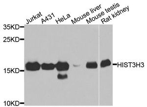
Western blot analysis of extracts of various cells, using Histone H3 antibody.Application
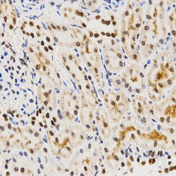
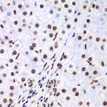
Immunohistochemistry of paraffin-embedded rat kidney tissue using Histone H3 antibody at dilution of 1:200 (x400 lens).Application
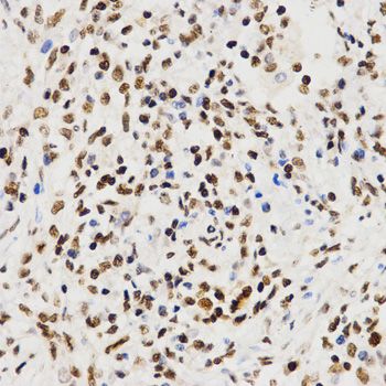
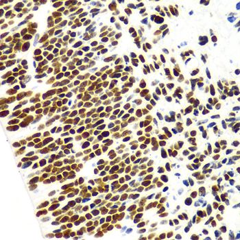
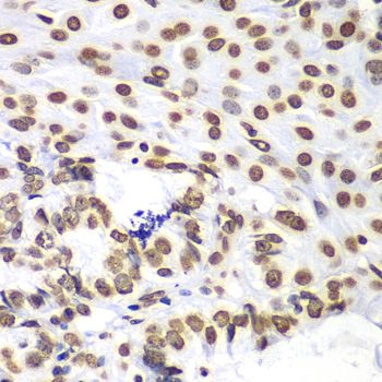
Immunohistochemistry of paraffin-embedded human lung cancer tissue using Histone H3 antibody at dilution of 1:200 (x400 lens).Application
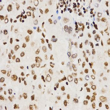
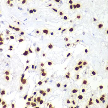
Immunohistochemistry of paraffin-embedded human liver cancer tissue using Histone H3 antibody at dilution of 1:200 (x400 lens).Application
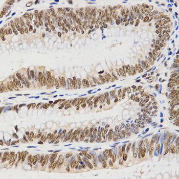
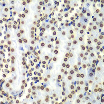
Immunohistochemistry of paraffin-embedded human rectal cancer tissue using Histone H3 antibody at dilution of 1:200 (x400 lens).Application
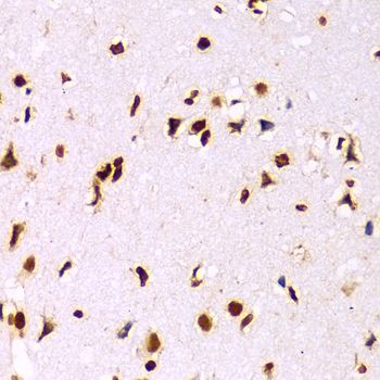
Immunohistochemistry of paraffin-embedded Human embryo brain using Histone H3 antibody at dilution of 1:100 (x400 lens).Application
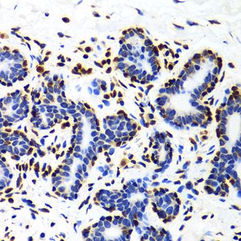
Immunohistochemistry of paraffin-embedded Human mammary gland using Histone H3 antibody at dilution of 1:100 (x400 lens).Application
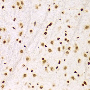
Immunohistochemistry of paraffin-embedded Mouse brain using Histone H3 antibody at dilution of 1:100 (x400 lens).Application
Immunohistochemistry of paraffin-embedded Rat brain using Histone H3 antibody at dilution of 1:100 (x400 lens).Application
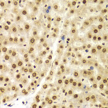
Immunohistochemistry of paraffin-embedded Rat liver using Histone H3 antibody at dilution of 1:100 (x400 lens).Application
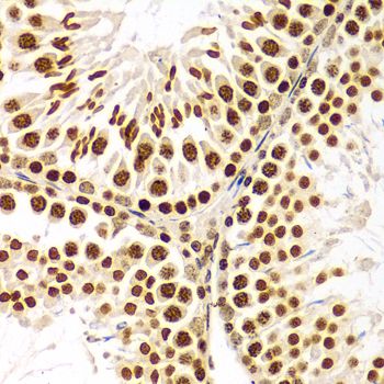
Immunohistochemistry of paraffin-embedded Rat testis using Histone H3 antibody at dilution of 1:100 (x400 lens).Application
Immunohistochemistry of paraffin-embedded rat kidney using HIST3H3 antibody at dilution of 1:100 (400x lens).Application
Immunohistochemistry of paraffin-embedded human liver cancer using HIST3H3 antibody at dilution of 1:100 (400x lens).Application
Immunohistochemistry of paraffin-embedded human oophoroma using HIST3H3 antibody at dilution of 1:100 (400x lens).Application
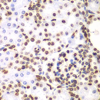
Immunohistochemistry of paraffin-embedded human kidney cancer using HIST3H3 antibody at dilution of 1:100 (400x lens).Application
Immunohistochemistry of paraffin-embedded mouse kidney using HIST3H3 antibody at dilution of 1:100 (400x lens).Application
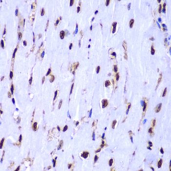
Immunohistochemistry of paraffin-embedded mouse heart using HIST3H3 antibody at dilution of 1:100 (400x lens).Application
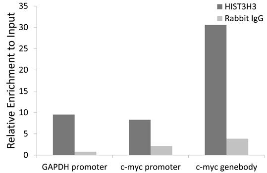
Chromatin Immunoprecipitation analysis extracts of HeLa cell line, using HIST3H3 antibody and rabbit IgG. The amount of immunoprecipitated DNA was checked by quantitative PCR. Histogram was constructed by the ratios of the immunoprecipitated DNA to the input.WB
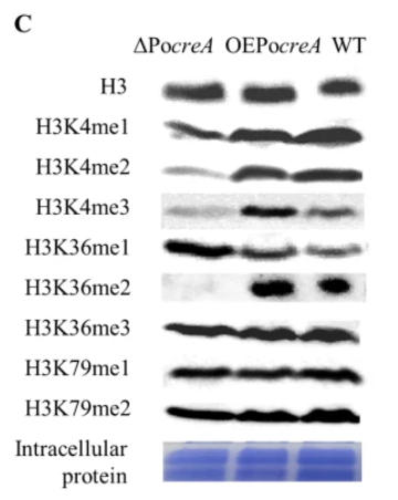
C Assays of histone methylation patterns using Western blot. Histone H3 was used as the loading control.WB
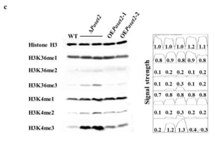
c Analysis of H3K4 and H3K36 methylation patterns through Western blot. Histone H3 was used as the loading control. The peak area and the data were obtained by the software Image J, representing the relative signal strength of the corresponding Western blot bands.WB
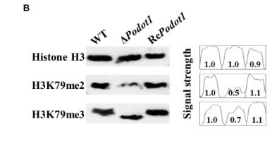
(B) Assay of the global histone methylation patterns at H3K79 in WT and ΔPodot1 through Western blot. Histone H3 was used as the loading control.WB
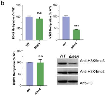
b Global methylation levels of H3K4, H3K9, and H3K27 in histone protein extracts from strain WT and ΔlaeA when grown on glucose for 24 h. Western blot assay of extracted histone of WT and ΔlaeA strains, which were carried out with specific antibodies for histone H3, H3K4me3, and H3K9me3.WB
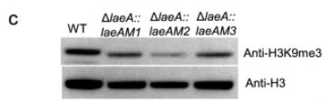
c Western blot assay of extracted histone protein from strains WT, ΔlaeA::LaeAM1, ΔlaeA::LaeAM2, and ΔlaeA::LaeAM3, with a specific antibody for H3K9me3.| Product Name | Histone H3 antibody |
|---|---|
| Antibody Type | Primary Antibodies |
| Product description | Rabbit Polyclonal antibody to Histone H3Histones are basic nuclear proteins that are responsible for the nucleosome structure of the chromosomal fiber in eukaryotes. Nucleosomes consist of approximately 146 bp of DNA wrapped around a histone octamer composed of pairs of each of the four core histones (H2A, H2B, H3, and H4). The chromatin fiber is further compacted through the interaction of a linker histone, H1, with the DNA between the nucleosomes to form higher order chromatin structures. This gene is intronless and encodes a member of the histone H3 family. Transcripts from this gene lack polyA tails; instead, they contain a palindromic termination element. This gene is located separately from the other H3 genes that are in the histone gene cluster on chromosome 6p22-p21.3. [provided by RefSeq, Jul 2008] |
| Immunogen | A synthetic peptide of human HIST3H3 |
| Clonality | Polyclonal |
|---|---|
| Isotype | IgG |
| Host Species | Rabbit |
| WB : 1:500 - 1:2000, IHC-P : 1:50 - 1:200, ICC/IF : 1:50 - 1:200, IP 1:50 - 1:200, ChIP 1:50 - 1:200. | |
| Concentration | 1mg/ml |
| Purification | Affinity purified |
| Gene Synonyms | H3.4 , H3t , HIST3H3 , histone cluster 3, H3 , H3FT , H3/g |
|---|---|
| Target | Histone H3 |
Application
Western blot analysis of extracts of various cells, using Histone H3 antibody.
Application
Immunohistochemistry of paraffin-embedded rat kidney tissue using Histone H3 antibody at dilution of 1:200 (x400 lens).
Application
Immunohistochemistry of paraffin-embedded human lung cancer tissue using Histone H3 antibody at dilution of 1:200 (x400 lens).
Application
Immunohistochemistry of paraffin-embedded human liver cancer tissue using Histone H3 antibody at dilution of 1:200 (x400 lens).
Application
Immunohistochemistry of paraffin-embedded human rectal cancer tissue using Histone H3 antibody at dilution of 1:200 (x400 lens).
Application
Immunohistochemistry of paraffin-embedded Human embryo brain using Histone H3 antibody at dilution of 1:100 (x400 lens).
Application
Immunohistochemistry of paraffin-embedded Human mammary gland using Histone H3 antibody at dilution of 1:100 (x400 lens).
Application
Immunohistochemistry of paraffin-embedded Mouse brain using Histone H3 antibody at dilution of 1:100 (x400 lens).
Application
Immunohistochemistry of paraffin-embedded Rat brain using Histone H3 antibody at dilution of 1:100 (x400 lens).
Application
Immunohistochemistry of paraffin-embedded Rat liver using Histone H3 antibody at dilution of 1:100 (x400 lens).
Application
Immunohistochemistry of paraffin-embedded Rat testis using Histone H3 antibody at dilution of 1:100 (x400 lens).
Application
Immunohistochemistry of paraffin-embedded rat kidney using HIST3H3 antibody at dilution of 1:100 (400x lens).
Application
Immunohistochemistry of paraffin-embedded human liver cancer using HIST3H3 antibody at dilution of 1:100 (400x lens).
Application
Immunohistochemistry of paraffin-embedded human oophoroma using HIST3H3 antibody at dilution of 1:100 (400x lens).
Application
Immunohistochemistry of paraffin-embedded human kidney cancer using HIST3H3 antibody at dilution of 1:100 (400x lens).
Application
Immunohistochemistry of paraffin-embedded mouse kidney using HIST3H3 antibody at dilution of 1:100 (400x lens).
Application
Immunohistochemistry of paraffin-embedded mouse heart using HIST3H3 antibody at dilution of 1:100 (400x lens).
Application
Chromatin Immunoprecipitation analysis extracts of HeLa cell line, using HIST3H3 antibody and rabbit IgG. The amount of immunoprecipitated DNA was checked by quantitative PCR. Histogram was constructed by the ratios of the immunoprecipitated DNA to the input.
WB
C Assays of histone methylation patterns using Western blot. Histone H3 was used as the loading control.
WB
c Analysis of H3K4 and H3K36 methylation patterns through Western blot. Histone H3 was used as the loading control. The peak area and the data were obtained by the software Image J, representing the relative signal strength of the corresponding Western blot bands.
WB
(B) Assay of the global histone methylation patterns at H3K79 in WT and ΔPodot1 through Western blot. Histone H3 was used as the loading control.
WB
b Global methylation levels of H3K4, H3K9, and H3K27 in histone protein extracts from strain WT and ΔlaeA when grown on glucose for 24 h. Western blot assay of extracted histone of WT and ΔlaeA strains, which were carried out with specific antibodies for histone H3, H3K4me3, and H3K9me3.
WB
c Western blot assay of extracted histone protein from strains WT, ΔlaeA::LaeAM1, ΔlaeA::LaeAM2, and ΔlaeA::LaeAM3, with a specific antibody for H3K9me3.| Application Notes | WB : 1:500 - 1:2000, IHC-P : 1:50 - 1:200, ICC/IF : 1:50 - 1:200, IP 1:50 - 1:200, ChIP 1:50 - 1:200. |
|---|
| Form | Liquid |
|---|---|
| Storage Instructions | Store at -20℃. Avoid freeze / thaw cycles. |
| Storage Buffer | PBS with 0.02% sodium azide, 50% glycerol, pH7.3. |


