Application
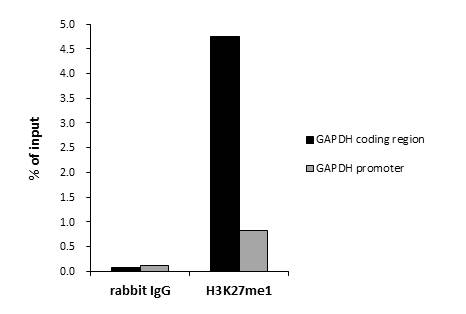
ChIP was performed with HeLa nuclear extract and either 2 μg of H3K27me1 antibody or control rabbit IgG. The precipitated DNA was detected by QPCR with primer set targeting to GAPDH coding region or GAPDH promoter.Application
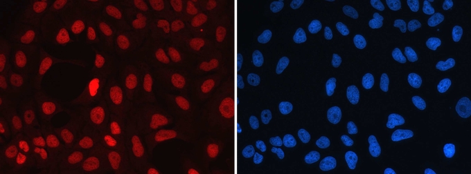
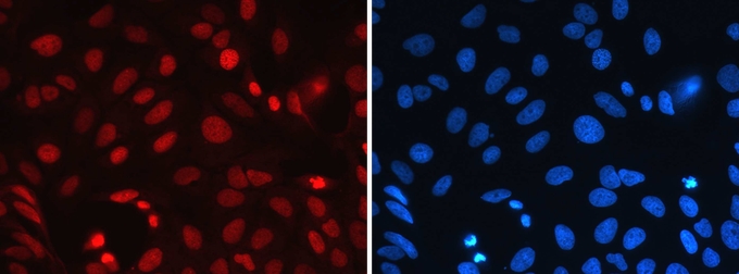
ICC/IF analysis of U2OS cells uaing H3K27me1 antibody and DAPI. Cells were fixed with 4% formaldehyde for 20’ and blocked with PBS/TX-100 containing 5% normal goat serum. The cells were immunofluorescently labeled with the H3K27me1 antibody at a dilution of 1:1,000 (red). The nuclei were stained with DAPI (blue).Application
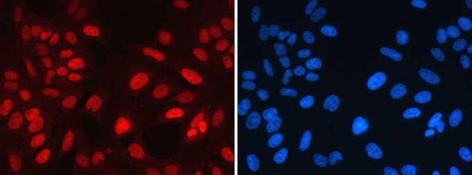
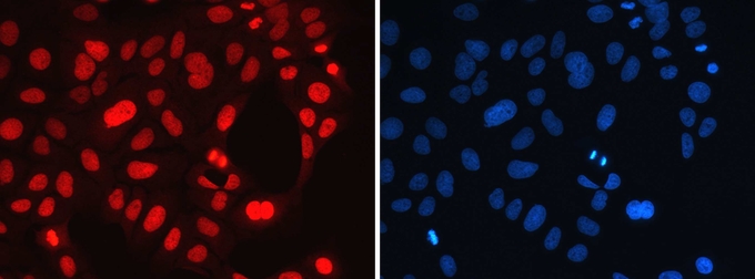
ICC/IF analysis of U2OS cells uaing H3K27 unmodified peptide-blocked H3K27me1 antibody and DAPI. Cells were fixed with 4% formaldehyde for 20’ and blocked with PBS/TX-100 containing 5% normal goat serum. The cells were immunofluorescently labeled with the H3K27 unmodified peptide-blocked H3K27me1 antibody at a dilution of 1:1,000 (red). The nuclei were stained with DAPI (blue).Application
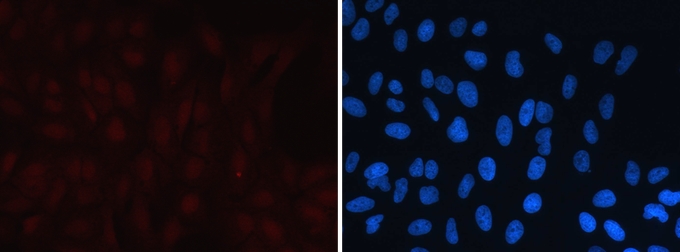
ICC/IF analysis of U2OS cells uaing H3K27me1 peptide-blocked H3K27me1 antibody and DAPI. Cells were fixed with 4% formaldehyde for 20’ and blocked with PBS/TX-100 containing 5% normal goat serum. The cells were immunofluorescently labeled with the H3K27me1 peptide-blocked H3K27me1 antibody at a dilution of 1:1,000 (red). The nuclei were stained with DAPI (blue).Application
ICC/IF analysis of U2OS cells uaing H3K27me2 peptide-blocked H3K27me1 antibody and DAPI. Cells were fixed with 4% formaldehyde for 20’ and blocked with PBS/TX-100 containing 5% normal goat serum. The cells were immunofluorescently labeled with the H3K27me2 peptide-blocked H3K27me1 antibody at a dilution of 1:1,000 (red). The nuclei were stained with DAPI (blue).Application
ICC/IF analysis of U2OS cells uaing H3K27me3 peptide-blocked H3K27me1 antibody and DAPI. Cells were fixed with 4% formaldehyde for 20’ and blocked with PBS/TX-100 containing 5% normal goat serum. The cells were immunofluorescently labeled with the H3K27me3 peptide-blocked H3K27me1 antibody at a dilution of 1:1,000 (red). The nuclei were stained with DAPI (blue).Application
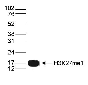
WB analysis of histone extracts (15 μg) from HeLa cells using H3K27me1 antibody diluted 1:1,000 in TBS-Tween containing 5% skimmed milk.Application
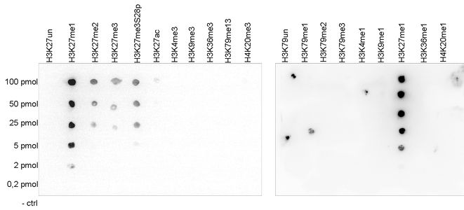
Dot blot analysis of peptides containing modified and unmodified sequences of histone H3 and H4 using H3K27me1 antibody at a dilution of 1:20,000. One hundred to 0.2 pmol of the peptide containing the respective histone modification were spotted on a membrane for analysis.Application
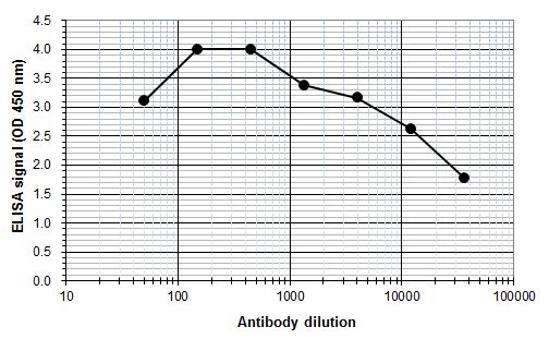
ELISA was performed using a serial dilution of Histone H3K27me1 (mono-Methyl Lys27) antibody in antigen coated wells. By plotting the absorbance against the antibody dilution, the titer of the purified antibody was estimated to be 1:32,900.| Product Name | Histone H3K27me1 (monomethyl Lys27) antibody - ChIP grade |
|---|---|
| Antibody Type | Primary Antibodies |
| Product description | Rabbit polyclonal antibody to Histone H3K27me1 (monomethyl Lys27) |
| Modification | monomethyl Lys27 |
| Clonality | Polyclonal |
|---|---|
| Host Species | Rabbit |
| Tested Applications | Dot,ELISA,ICC/IF,WB,ChIP |
| ChIP (2 µg/ChIP), ELISA (1:100 - 1:500), Dot blotting (1:20,000), Western blotting (1:1,000) | |
| Species Reactivity | Human |
| Concentration | 1mg/ml |
| Purification | Affinity purified |
| Gene Synonyms | mono Methyl Histone H3 Lys27, mono Methyl K27 Histone H3, histone cluster 1, H3a, mono-Methyl K27 Histone H3, Histone H3 mono-Methyl Lys27, H3/A, HIST1H3A, mono Methyl Histone H3, mono Methyl Histone H3 K27, monoMethyl K27 Histone H3, Histone H3 monoMethyl |
|---|---|
| Target | Histone H3 (mono-Methyl Lys27) |
Application
ChIP was performed with HeLa nuclear extract and either 2 μg of H3K27me1 antibody or control rabbit IgG. The precipitated DNA was detected by QPCR with primer set targeting to GAPDH coding region or GAPDH promoter.
Application
ICC/IF analysis of U2OS cells uaing H3K27me1 antibody and DAPI. Cells were fixed with 4% formaldehyde for 20’ and blocked with PBS/TX-100 containing 5% normal goat serum. The cells were immunofluorescently labeled with the H3K27me1 antibody at a dilution of 1:1,000 (red). The nuclei were stained with DAPI (blue).
Application
ICC/IF analysis of U2OS cells uaing H3K27 unmodified peptide-blocked H3K27me1 antibody and DAPI. Cells were fixed with 4% formaldehyde for 20’ and blocked with PBS/TX-100 containing 5% normal goat serum. The cells were immunofluorescently labeled with the H3K27 unmodified peptide-blocked H3K27me1 antibody at a dilution of 1:1,000 (red). The nuclei were stained with DAPI (blue).
Application
ICC/IF analysis of U2OS cells uaing H3K27me1 peptide-blocked H3K27me1 antibody and DAPI. Cells were fixed with 4% formaldehyde for 20’ and blocked with PBS/TX-100 containing 5% normal goat serum. The cells were immunofluorescently labeled with the H3K27me1 peptide-blocked H3K27me1 antibody at a dilution of 1:1,000 (red). The nuclei were stained with DAPI (blue).
Application
ICC/IF analysis of U2OS cells uaing H3K27me2 peptide-blocked H3K27me1 antibody and DAPI. Cells were fixed with 4% formaldehyde for 20’ and blocked with PBS/TX-100 containing 5% normal goat serum. The cells were immunofluorescently labeled with the H3K27me2 peptide-blocked H3K27me1 antibody at a dilution of 1:1,000 (red). The nuclei were stained with DAPI (blue).
Application
ICC/IF analysis of U2OS cells uaing H3K27me3 peptide-blocked H3K27me1 antibody and DAPI. Cells were fixed with 4% formaldehyde for 20’ and blocked with PBS/TX-100 containing 5% normal goat serum. The cells were immunofluorescently labeled with the H3K27me3 peptide-blocked H3K27me1 antibody at a dilution of 1:1,000 (red). The nuclei were stained with DAPI (blue).
Application
WB analysis of histone extracts (15 μg) from HeLa cells using H3K27me1 antibody diluted 1:1,000 in TBS-Tween containing 5% skimmed milk.
Application
Dot blot analysis of peptides containing modified and unmodified sequences of histone H3 and H4 using H3K27me1 antibody at a dilution of 1:20,000. One hundred to 0.2 pmol of the peptide containing the respective histone modification were spotted on a membrane for analysis.
Application
ELISA was performed using a serial dilution of Histone H3K27me1 (mono-Methyl Lys27) antibody in antigen coated wells. By plotting the absorbance against the antibody dilution, the titer of the purified antibody was estimated to be 1:32,900.| Application Notes | ChIP (2 µg/ChIP), ELISA (1:100 - 1:500), Dot blotting (1:20,000), Western blotting (1:1,000) |
|---|
| Form | Liquid |
|---|---|
| Storage Instructions | Store at -20°C; for long storage, store at -80°C. Avoid multiple freeze-thaw cycles. |
| Storage Buffer | PBS containing 0.05% azide and 0.05% ProClin 300. |


