Application
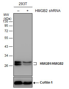
Non-transfected (–) and transfected (+) 293T whole cell extracts (30 µg) were separated by 12% SDS-PAGE, and the membrane was blotted with HMGB1/HMGB2 antibody diluted at 1:20000.Application
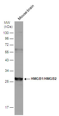
Mouse tissue extract (50 µg) was separated by 12% SDS-PAGE, and the membrane was blotted with HMGB1/HMGB2 antibody diluted at 1:1000.Application
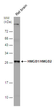
Rat tissue extract (50 µg) was separated by 12% SDS-PAGE, and the membrane was blotted with HMGB1/HMGB2 antibody diluted at 1:2000.Application
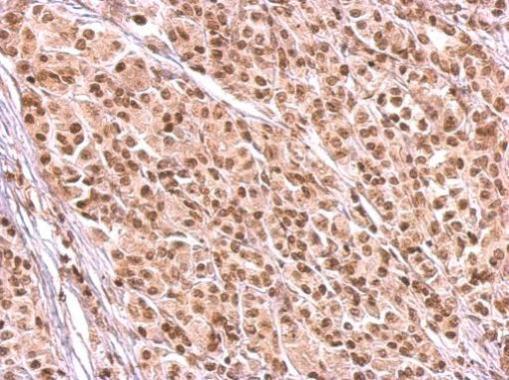
HMGB1/HMGB2 antibody detects HMGB2 protein at nucleus on human duodenal adenocarcinoma tissue by immunohistochemical analysis. Sample: Paraffin-embedded human duodenal adenocarcinoma tissue. HMGB1/HMGB2 antibody dilution: 1:500.Application
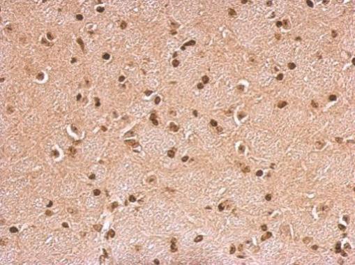
HMGB1/HMGB2 antibody detects HMGB2 protein at cytosol on mouse brain by immunohistochemical analysis. Sample: Paraffin-embedded mouse brain. HMGB1/HMGB2 antibody dilution: 1:500.Application
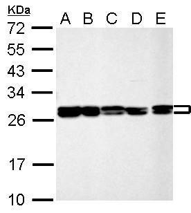
HMGB1/HMGB2 antibody detects HMGB2 protein by Western blot analysis. A. 30 µg 293T whole cell lysate/extract B. 30 µg A431 whole cell lysate/extract C. 30 µg HeLa whole cell lysate/extract D. 30 µg HepG2 whole cell lysate/extract E. 30 µg A375 whole cell lysate/extract 12 % SDS-PAGE HMGB1/HMGB2 antibody dilution: 1:10000Application
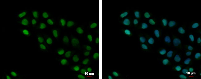
HMGB1/HMGB2 antibody detects HMGB1/HMGB2 protein at nucleus by immunofluorescent analysis. Sample: A375 cells were fixed in 4% paraformaldehyde at RT for 15 min. Green: HMGB1/HMGB2 protein stained by HMGB1/HMGB2 antibody diluted at 1:500. Blue: Hoechst 33342 staining. Scale bar = 10 μm.Application
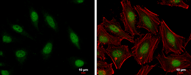
HMGB1/HMGB2 antibody detects HMGB1/HMGB2 protein at nucleus by immunofluorescent analysis. Sample: HeLa cells were fixed in 4% paraformaldehyde at RT for 15 min. Green: HMGB1/HMGB2 protein stained by HMGB1/HMGB2 antibody diluted at 1:2000. Red: phalloidin, a cytoskeleton marker, diluted at 1:200. Blue: Hoechst 33342 staining. Scale bar = 10 μm.| Product Name | HMGB1/HMGB2 antibody |
|---|---|
| Antibody Type | Primary Antibodies |
| Product description | Rabbit Polyclonal antibody to HMGB1/HMGB2 (high-mobility group box 2)This gene encodes a member of the non-histone chromosomal high mobility group protein family. The proteins of this family are chromatin-associated and ubiquitously distributed in the nucleus of higher eukaryotic cells. In vitro studies have demonstrated that this protein is able to efficiently bend DNA and form DNA circles. These studies suggest a role in facilitating cooperative interactions between cis-acting proteins by promoting DNA flexibility. This protein was also reported to be involved in the final ligation step in DNA end-joining processes of DNA double-strand breaks repair and V(D)J recombination. [provided by RefSeq] |
| Immunogen | Full length human HMGB1/HMGB2 Recombinant protein. |
| Clonality | Polyclonal |
|---|---|
| Isotype | IgG |
| Host Species | Rabbit |
| Tested Applications | ICC/IFIHC-PWB |
| *Optimal dilutions/concentrations should be determined by the researcher.
: |
|
| Species Reactivity | HumanMouseRat |
| Predicted Reactivity | Pig, Chicken, BovinePig (99%), Chicken (82%), Bovine (100%) |
| Concentration | 1mg/ml (Please refer to the vial label for the specific concentration) |
| Purification | Affinity purified |
| Gene Synonyms | HMG2 antibody HMGB2 antibody high mobility group protein 2 antibody high-mobility group (nonhistone chromosomal) protein 2 antibody high mobility group protein B2 antibody HMG-2 antibody high-mobility group box 2 antibody |
|---|---|
| Molecular Weight(MW) | 24 kDa (note) (3) |
| Cellular Localization | Nucleus |
Application
Non-transfected (–) and transfected (+) 293T whole cell extracts (30 µg) were separated by 12% SDS-PAGE, and the membrane was blotted with HMGB1/HMGB2 antibody diluted at 1:20000.
Application
Mouse tissue extract (50 µg) was separated by 12% SDS-PAGE, and the membrane was blotted with HMGB1/HMGB2 antibody diluted at 1:1000.
Application
Rat tissue extract (50 µg) was separated by 12% SDS-PAGE, and the membrane was blotted with HMGB1/HMGB2 antibody diluted at 1:2000.
Application
HMGB1/HMGB2 antibody detects HMGB2 protein at nucleus on human duodenal adenocarcinoma tissue by immunohistochemical analysis. Sample: Paraffin-embedded human duodenal adenocarcinoma tissue. HMGB1/HMGB2 antibody dilution: 1:500.
Application
HMGB1/HMGB2 antibody detects HMGB2 protein at cytosol on mouse brain by immunohistochemical analysis. Sample: Paraffin-embedded mouse brain. HMGB1/HMGB2 antibody dilution: 1:500.
Application
HMGB1/HMGB2 antibody detects HMGB2 protein by Western blot analysis. A. 30 µg 293T whole cell lysate/extract B. 30 µg A431 whole cell lysate/extract C. 30 µg HeLa whole cell lysate/extract D. 30 µg HepG2 whole cell lysate/extract E. 30 µg A375 whole cell lysate/extract 12 % SDS-PAGE HMGB1/HMGB2 antibody dilution: 1:10000
Application
HMGB1/HMGB2 antibody detects HMGB1/HMGB2 protein at nucleus by immunofluorescent analysis. Sample: A375 cells were fixed in 4% paraformaldehyde at RT for 15 min. Green: HMGB1/HMGB2 protein stained by HMGB1/HMGB2 antibody diluted at 1:500. Blue: Hoechst 33342 staining. Scale bar = 10 μm.
Application
HMGB1/HMGB2 antibody detects HMGB1/HMGB2 protein at nucleus by immunofluorescent analysis. Sample: HeLa cells were fixed in 4% paraformaldehyde at RT for 15 min. Green: HMGB1/HMGB2 protein stained by HMGB1/HMGB2 antibody diluted at 1:2000. Red: phalloidin, a cytoskeleton marker, diluted at 1:200. Blue: Hoechst 33342 staining. Scale bar = 10 μm.| Positive Control | 293T , A431 , HeLa , HepG2 , A375 , mouse brain , rat brain |
|---|---|
| Application Notes | *Optimal dilutions/concentrations should be determined by the researcher.
: |
| Form | Liquid |
|---|---|
| Storage Instructions | Keep as concentrated solution. Aliquot and store at -20ºC or below. Avoid multiple freeze-thaw cycles. |
| Storage Buffer | 1XPBS, 20% Glycerol (pH7). 0.01% Thimerosal was added as a preservative. |


