Application
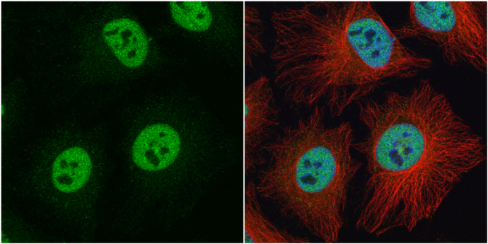
hnRNP C1/C2 antibody detects hnRNP C1/C2 protein at nucleus by immunofluorescent analysis. Sample: HeLa cells were fixed in 4% paraformaldehyde at RT for 15 min. Green: hnRNP C1/C2 protein stained by hnRNP C1/C2 antibody diluted at 1:500. Red: alpha Tubulin, a cytoskeleton marker, stained by alpha Tubulin antibody [GT114] diluted at 1:1000. Blue: Hoechst 33342 staining.Application
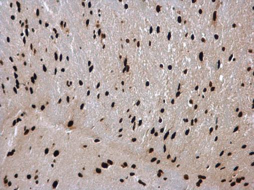
hnRNP C1/C2 antibody detects hnRNP C1/C2 protein at nucleus in mouse brain by immunohistochemical analysis. Sample: Paraffin-embedded mouse brain. hnRNP C1/C2 antibody diluted at 1:500.Application
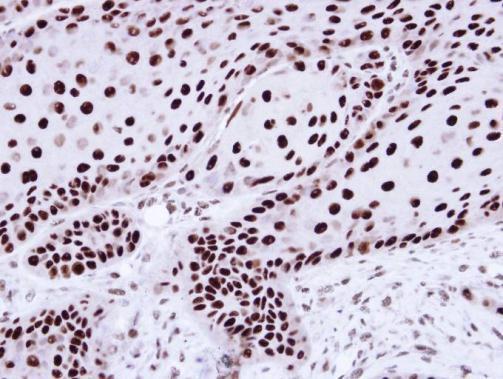
Immunohistochemical analysis of paraffin-embedded Cal27 Xenograft, using hnRNP C1/C2 antibody at 1:100 dilution.Application
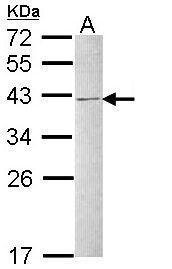
Sample (30 µg of whole cell lysate) A: Raji 12% SDS PAGE diluted at 1:1000 The HRP-conjugated anti-rabbit IgG antibody was used to detect the primary antibody.Application
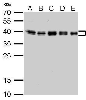
hnRNP C1/C2 antibody detects HNRNPC protein by western blot analysis. A. 30 µg Neuro2A whole cell lysate/extract B. 30 µg NIH-3T3 whole cell lysate/extract C. 30 µg BCL-1 whole cell lysate/extract D. 30 µg Raw264.7 whole cell lysate/extract E. 30 µg C2C12 whole cell lysate/extract 12% SDS-PAGE hnRNP C1/C2 antibody dilution: 1:1000 The HRP-conjugated anti-rabbit IgG antibody was used to detect the primary antibody.Application
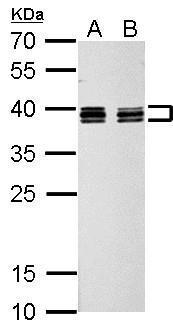
hnRNP C1/C2 antibody detects HNRNPC protein by western blot analysis. A. 30 µg PC-12 whole cell lysate/extract B. 30 µg Rat2 whole cell lysate/extract 12% SDS-PAGE hnRNP C1/C2 antibody dilution: 1:1000 The HRP-conjugated anti-rabbit IgG antibody was used to detect the primary antibody.| Product Name | hnRNP C1/C2 antibody |
|---|---|
| Antibody Type | Primary Antibodies |
| Product description | Rabbit Polyclonal antibody to hnRNP C1/C2 (heterogeneous nuclear ribonucleoprotein C (C1/C2))This gene belongs to the subfamily of ubiquitously expressed heterogeneous nuclear ribonucleoproteins (hnRNPs). The hnRNPs are RNA binding proteins and they complex with heterogeneous nuclear RNA (hnRNA). These proteins are associated with pre-mRNAs in the nucleus and appear to influence pre-mRNA processing and other aspects of mRNA metabolism and transport. While all of the hnRNPs are present in the nucleus, some seem to shuttle between the nucleus and the cytoplasm. The hnRNP proteins have distinct nucleic acid binding properties. The protein encoded by this gene can act as a tetramer and is involved in the assembly of 40S hnRNP particles. Multiple transcript variants encoding at least two different isoforms have been described for this gene. [provided by RefSeq] |
| Immunogen | Recombinant protein encompassing a sequence within the center region of human hnRNP C1/C2. The exact sequence is proprietary. |
| Clonality | Polyclonal |
|---|---|
| Isotype | IgG |
| Host Species | Rabbit |
| Tested Applications | ICC/IFIHC-PIPWB |
| *Optimal dilutions/concentrations should be determined by the researcher.
: |
|
| Species Reactivity | HumanMouseRat |
| Predicted Reactivity | Xenopus laevis, Pig, Rabbit, Rhesus Monkey, Chimpanzee, BovineXenopus laevis (88%), Pig (100%), Rabbit (100%), Rhesus Monkey (100%), Chimpanzee (100%), Bovine (100%) |
| Concentration | 1mg/ml |
| Purification | Affinity purified |
| Gene Synonyms | C1 antibody C2 antibody HNRNP antibody HNRPC antibody MGC104306 antibody MGC105117 antibody MGC117353 antibody MGC131677 antibody SNRPC antibody HNRNPC antibody hnRNP C1/C2 antibody nuclear ribonucleoprotein particle C1 protein antibody heterog |
|---|---|
| Molecular Weight(MW) | 34 kDa (note) (3) |
| Cellular Localization | Nucleus |
Application
hnRNP C1/C2 antibody detects hnRNP C1/C2 protein at nucleus by immunofluorescent analysis. Sample: HeLa cells were fixed in 4% paraformaldehyde at RT for 15 min. Green: hnRNP C1/C2 protein stained by hnRNP C1/C2 antibody diluted at 1:500. Red: alpha Tubulin, a cytoskeleton marker, stained by alpha Tubulin antibody [GT114] diluted at 1:1000. Blue: Hoechst 33342 staining.
Application
hnRNP C1/C2 antibody detects hnRNP C1/C2 protein at nucleus in mouse brain by immunohistochemical analysis. Sample: Paraffin-embedded mouse brain. hnRNP C1/C2 antibody diluted at 1:500.
Application
Immunohistochemical analysis of paraffin-embedded Cal27 Xenograft, using hnRNP C1/C2 antibody at 1:100 dilution.
Application
Sample (30 µg of whole cell lysate) A: Raji 12% SDS PAGE diluted at 1:1000 The HRP-conjugated anti-rabbit IgG antibody was used to detect the primary antibody.
Application
hnRNP C1/C2 antibody detects HNRNPC protein by western blot analysis. A. 30 µg Neuro2A whole cell lysate/extract B. 30 µg NIH-3T3 whole cell lysate/extract C. 30 µg BCL-1 whole cell lysate/extract D. 30 µg Raw264.7 whole cell lysate/extract E. 30 µg C2C12 whole cell lysate/extract 12% SDS-PAGE hnRNP C1/C2 antibody dilution: 1:1000 The HRP-conjugated anti-rabbit IgG antibody was used to detect the primary antibody.
Application
hnRNP C1/C2 antibody detects HNRNPC protein by western blot analysis. A. 30 µg PC-12 whole cell lysate/extract B. 30 µg Rat2 whole cell lysate/extract 12% SDS-PAGE hnRNP C1/C2 antibody dilution: 1:1000 The HRP-conjugated anti-rabbit IgG antibody was used to detect the primary antibody.| Positive Control | Raji , Neuro2A , NIH-3T3 , BCL-1 , Raw264.7 , C2C12 , PC-12 , Rat2 |
|---|---|
| Application Notes | *Optimal dilutions/concentrations should be determined by the researcher.
: |
| Form | Liquid |
|---|---|
| Storage Instructions | Keep as concentrated solution. Aliquot and store at -20ºC or below. Avoid multiple freeze-thaw cycles. |
| Storage Buffer | 0.1M Tris, 0.1M Glycine, 20% Glycerol (pH7). 0.01% Thimerosal was added as a preservative. |


