Application
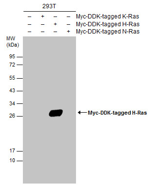
Non-transfected (–) and transfected (+) 293T whole cell extracts (30 µg) were separated by 12% SDS-PAGE, and the membrane was blotted with H-Ras antibody diluted at 1:5000. The HRP-conjugated anti-rabbit IgG antibody was used to detect the primary antibody.Application
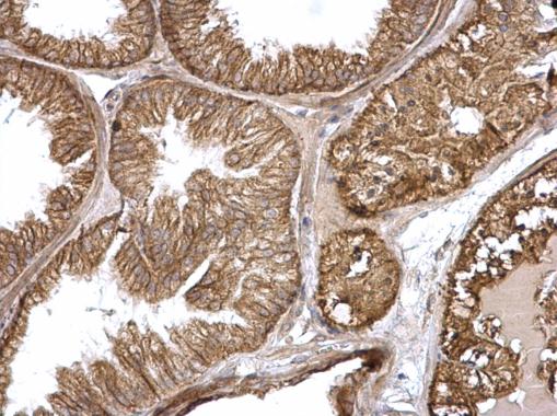
H-Ras antibody detects H-Ras protein at membrane on mouse prostate by immunohistochemical analysis. Sample: Paraffin-embedded mouse prostate. H-Ras antibody dilution: 1:500.Application
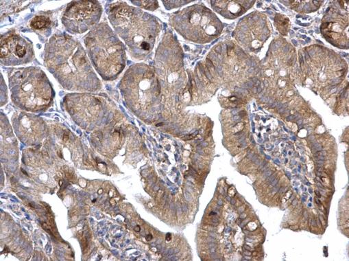
H-Ras antibody detects H-Ras protein at membrane on mouse intestine by immunohistochemical analysis. Sample: Paraffin-embedded mouse intestine. H-Ras antibody dilution: 1:500.Application
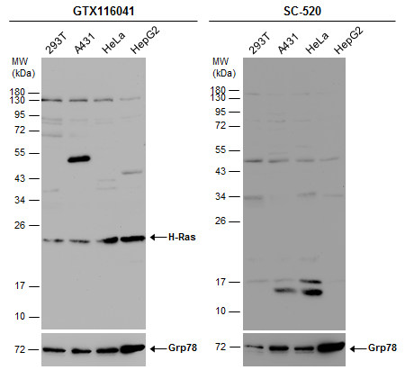
Various whole cell extracts (30 µg) were separated by 12% SDS-PAGE, and the membranes were blotted with H-Ras antibody diluted at 1:500 and competitor's antibody (SC-520) diluted by 1:200. The HRP-conjugated anti-rabbit IgG antibody was used to detect the primary antibody.Application
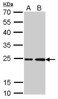
H-Ras antibody detects HRAS protein by western blot analysis. A. 30 µg PC-12 whole cell lysate/extract B. 30 µg Rat2 whole cell lysate/extract 12% SDS-PAGE H-Ras antibody dilution: 1:1000 The HRP-conjugated anti-rabbit IgG antibody was used to detect the primary antibody.Application
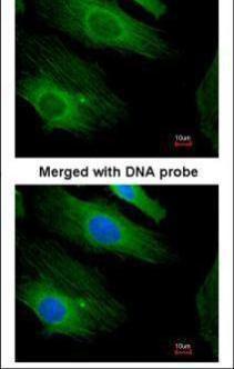
Immunofluorescence analysis of paraformaldehyde-fixed HeLa, using H-Ras antibody at 1:200 dilution.Application
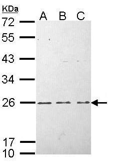
Sample (30 µg of whole cell lysate) A: HeLa B: HepG2 C: Molt-4 12% SDS PAGE diluted at 1:1000 The HRP-conjugated anti-rabbit IgG antibody was used to detect the primary antibody.| Product Name | H-Ras antibody |
|---|---|
| Antibody Type | Primary Antibodies |
| Product description | Rabbit Polyclonal antibody to H-Ras (v-Ha-ras Harvey rat sarcoma viral oncogene homolog)This gene belongs to the Ras oncogene family, whose members are related to the transforming genes of mammalian sarcoma retroviruses. The products encoded by these genes function in signal transduction pathways. These proteins can bind GTP and GDP, and they have intrinsic GTPase activity. This protein undergoes a continuous cycle of de- and re-palmitoylation, which regulates its rapid exchange between the plasma membrane and the Golgi apparatus. Mutations in this gene cause Costello syndrome, a disease characterized by increased growth at the prenatal stage, growth deficiency at the postnatal stage, predisposition to tumor formation, mental retardation, skin and musculoskeletal abnormalities, distinctive facial appearance and cardiovascular abnormalities. Defects in this gene are implicated in a variety of cancers, including bladder cancer, follicular thyroid cancer, and oral squamous cell carcinoma. Multiple transcript variants, which encode different isoforms, have been identified for this gene. [provided by RefSeq] |
| Immunogen | Carrier-protein conjugated synthetic peptide encompassing a sequence within the C-terminus region of human H-Ras. The exact sequence is proprietary. |
| Clonality | Polyclonal |
|---|---|
| Isotype | IgG |
| Host Species | Rabbit |
| Tested Applications | ICC/IFIHC-PWB |
| *Optimal dilutions/concentrations should be determined by the researcher.
: |
|
| Species Reactivity | HumanMouseRat |
| Predicted Reactivity | Xenopus laevis, Chicken, Rhesus MonkeyXenopus laevis (100%), Chicken (100%), Rhesus Monkey (100%) |
| Concentration | 1mg/ml (Please refer to the vial label for the specific concentration) |
| Purification | Affinity purified |
| Gene Synonyms | C-BAS/HAS antibody C-H-RAS antibody C-HA-RAS1 antibody CTLO antibody H-RASIDX antibody HAMSV antibody HRAS1 antibody K-RAS antibody N-RAS antibody RASH1 antibody HRAS antibody transforming protein p21 antibody Ha-Ras1 proto-oncoprotein antibody |
|---|---|
| Molecular Weight(MW) | 21 kDa (note) (3) |
| Tissue Specificity | This antibody is specific for H-Ras protein, and it does not cross react with human N-Ras or K-Ras protein. |
| Cellular Localization | Cell membrane; Lipid-anchor; Cytoplasmic side , Golgi apparatus membrane; Lipid-anchor |
Application
Non-transfected (–) and transfected (+) 293T whole cell extracts (30 µg) were separated by 12% SDS-PAGE, and the membrane was blotted with H-Ras antibody diluted at 1:5000. The HRP-conjugated anti-rabbit IgG antibody was used to detect the primary antibody.
Application
H-Ras antibody detects H-Ras protein at membrane on mouse prostate by immunohistochemical analysis. Sample: Paraffin-embedded mouse prostate. H-Ras antibody dilution: 1:500.
Application
H-Ras antibody detects H-Ras protein at membrane on mouse intestine by immunohistochemical analysis. Sample: Paraffin-embedded mouse intestine. H-Ras antibody dilution: 1:500.
Application
Various whole cell extracts (30 µg) were separated by 12% SDS-PAGE, and the membranes were blotted with H-Ras antibody diluted at 1:500 and competitor's antibody (SC-520) diluted by 1:200. The HRP-conjugated anti-rabbit IgG antibody was used to detect the primary antibody.
Application
H-Ras antibody detects HRAS protein by western blot analysis. A. 30 µg PC-12 whole cell lysate/extract B. 30 µg Rat2 whole cell lysate/extract 12% SDS-PAGE H-Ras antibody dilution: 1:1000 The HRP-conjugated anti-rabbit IgG antibody was used to detect the primary antibody.
Application
Immunofluorescence analysis of paraformaldehyde-fixed HeLa, using H-Ras antibody at 1:200 dilution.
Application
Sample (30 µg of whole cell lysate) A: HeLa B: HepG2 C: Molt-4 12% SDS PAGE diluted at 1:1000 The HRP-conjugated anti-rabbit IgG antibody was used to detect the primary antibody.| Positive Control | 293T , A431 , HeLa , HepG2 , Molt-4 , 293T transfected with H-Ras , PC-12 , Rat2 , *RFL6 , *PLC5Rat2 , *RFL6 , *PLC5 |
|---|---|
| Application Notes | *Optimal dilutions/concentrations should be determined by the researcher.
: |
| Form | Liquid |
|---|---|
| Storage Instructions | Keep as concentrated solution. Aliquot and store at -20ºC or below. Avoid multiple freeze-thaw cycles. |
| Storage Buffer | 0.1M Tris, 0.1M Glycine, 20% Glycerol (pH7). 0.01% Thimerosal was added as a preservative. |


