Application
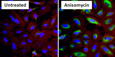
Immunofluorescent analysis of Phospho Heat Shock Protein 27 (p-HSP27) pSer15 (green) in HeLa cells either left untreated (left panel) or treated with 10uM Anisomysin (right panel) for 30 minutes. Formalin fixed cells were permeabilized with 0.1% Triton X-100 in TBS for 10 minutes at room temperature and blocked with 1% Blocker BSA for 15 minutes at room temperature. Cells were probed with a p-HSP27 pSer15 polyclonal antibody at a dilution of 1:50 for at least 1 hour at room temperature, washed with PBS, and incubated with DyLight 488 goat anti-rabbit IgG secondary antibody at a dilution of 1:400 for 30 minutes at room temperature. F-Actin (red) was stained with DyLight 554 Phalloidin and nuclei (blue) were stained with Hoechst dye.Application
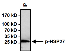
Immunoprecipitation of Phospho Heat Shock Protein 27 (p-HSP27) pSer15 was performed on HeLa cells treated with 10uM Anisomysin for 30 minutes. Antigen-antibody complexes were formed by incubating 500ug of whole cell lysate with 3ug of p-HSP27 pSer15 polyclonal antibody overnight on a rocking platform at 4°C. The immune complexes were captured on 50ul Protein A/G Agarose, washed extensively, and eluted. Samples were then resolved on a 4-20% Tris-HCl polyacrylamide gel, transferred to a PVDF membrane, and blocked with 5% BSA/TBST for at least 1 hour. The membrane was probed with a p-HSP27 pSer15 polyclonal antibody at a dilution of 1:500 overnight rotating at 4°C, washed with TBST, and probed with an IP detection reagent at a dilution of 1:1000 for at least 1 hour. Chemiluminescent detection was performedApplication
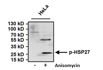
Western blot analysis of Phospho Heat Shock Protein 27 (p-HSP27) pSer15 was performed by loading 50ug of HeLa cell lysates from untreated cells (left lane) or cells treated with 10uM Anisomycin (right lane) for 30 minutes and 15ul onto a 4-20% Tris-HCl polyacrylamide gel. Proteins were transferred to a PVDF membrane and blocked with 5% BSA/TBST for at least 1 hour. The membrane was probed with a p-HSP27 pSer15 polyclonal antibody at a dilution of 1:500 overnight at 4°C on a rocking platform, washed with TBS-0.1%Tween-20, and probed with a goat anti-rabbit IgG-HRP secondary antibody at a dilution of 1:20,000 for at least 1 hour. Chemiluminescent detection was performed.| Product Name | HSP27 (phospho Ser15) antibody |
|---|---|
| Antibody Type | Primary Antibodies |
| Product description | Rabbit Polyclonal antibody to HSP27 (phospho Ser15)HSP27 also referred to as the Estrogen-Regulated 24K protein and HSP28, is one of several small heat shock proteins (HSP) produced by all organisms studied. HSP27 synthesis is induced by elevated temperature, as well as estrogen in hormone responsive cells. Interestingly, human HSP27 also shares greater than 50% homology with low molecular weight Drosophila HSP's and mammalian a-crystalline lens protein. Because of the estrogen responsive nature of HSP27, this protein has been studied extensively in human estrogen responsive tissues such as cervix, endometrium and breast tissue. This work has led to the suggestion that HSP27 may be a useful marker in classifying various hormone sensitive tumors. |
| Immunogen | Synthetic phosphopeptide corresponding to the residues L(10) L R G P (pS) W D P F R C(21) of human HSP27. |
| Modification | p-Ser15 |
| Clonality | Polyclonal |
|---|---|
| Host Species | Rabbit |
| Tested Applications | ICC/IFIHCIPWB |
| ICC/IF: 1:50 - 1:200: |
|
| Species Reactivity | HumanRatRabbit |
| Concentration | 1mg/ml (Please refer to the vial label for the specific concentration) |
| Purification | Affinity purified |
| Gene Synonyms | Hsp27 (phospho S15) Hsp27 (phospho Ser15) Hsp27 (pS15) Hsp27 (pSer15) Hsp27 phospho S15 Hsp27 phospho Ser15 Phospho Hsp27 (S15) Phospho Hsp27 (Ser15) Phospho Hsp27 (pS15) Phospho Hsp27 (pSer15) |
|---|---|
| Target | Phospho-Heat Shock Prot. 27 pSer15 |
Application
Immunofluorescent analysis of Phospho Heat Shock Protein 27 (p-HSP27) pSer15 (green) in HeLa cells either left untreated (left panel) or treated with 10uM Anisomysin (right panel) for 30 minutes. Formalin fixed cells were permeabilized with 0.1% Triton X-100 in TBS for 10 minutes at room temperature and blocked with 1% Blocker BSA for 15 minutes at room temperature. Cells were probed with a p-HSP27 pSer15 polyclonal antibody at a dilution of 1:50 for at least 1 hour at room temperature, washed with PBS, and incubated with DyLight 488 goat anti-rabbit IgG secondary antibody at a dilution of 1:400 for 30 minutes at room temperature. F-Actin (red) was stained with DyLight 554 Phalloidin and nuclei (blue) were stained with Hoechst dye.
Application
Immunoprecipitation of Phospho Heat Shock Protein 27 (p-HSP27) pSer15 was performed on HeLa cells treated with 10uM Anisomysin for 30 minutes. Antigen-antibody complexes were formed by incubating 500ug of whole cell lysate with 3ug of p-HSP27 pSer15 polyclonal antibody overnight on a rocking platform at 4°C. The immune complexes were captured on 50ul Protein A/G Agarose, washed extensively, and eluted. Samples were then resolved on a 4-20% Tris-HCl polyacrylamide gel, transferred to a PVDF membrane, and blocked with 5% BSA/TBST for at least 1 hour. The membrane was probed with a p-HSP27 pSer15 polyclonal antibody at a dilution of 1:500 overnight rotating at 4°C, washed with TBST, and probed with an IP detection reagent at a dilution of 1:1000 for at least 1 hour. Chemiluminescent detection was performed
Application
Western blot analysis of Phospho Heat Shock Protein 27 (p-HSP27) pSer15 was performed by loading 50ug of HeLa cell lysates from untreated cells (left lane) or cells treated with 10uM Anisomycin (right lane) for 30 minutes and 15ul onto a 4-20% Tris-HCl polyacrylamide gel. Proteins were transferred to a PVDF membrane and blocked with 5% BSA/TBST for at least 1 hour. The membrane was probed with a p-HSP27 pSer15 polyclonal antibody at a dilution of 1:500 overnight at 4°C on a rocking platform, washed with TBS-0.1%Tween-20, and probed with a goat anti-rabbit IgG-HRP secondary antibody at a dilution of 1:20,000 for at least 1 hour. Chemiluminescent detection was performed.| Application Notes | ICC/IF: 1:50 - 1:200: |
|---|
| Form | Liquid |
|---|---|
| Storage Instructions | Keep as concentrated solution. Aliquot and store at -20ºC or below. Avoid multiple freeze-thaw cycles. |
| Storage Buffer | PBS containing 1 mg/ml BSA and 0.05% sodium azide. |


