Application
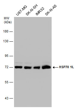
Various whole cell extracts (30 µg) were separated by 7.5% SDS-PAGE, and the membrane was blotted with HSP70 1L antibody [N1N3] diluted at 1:1000. The HRP-conjugated anti-rabbit IgG antibody was used to detect the primary antibody.Application
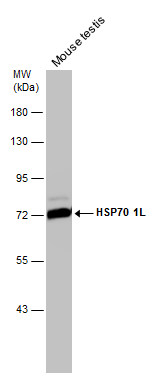
Mouse tissue extract (50 µg) was separated by 7.5% SDS-PAGE, and the membrane was blotted with HSP70 1L antibody [N1N3] diluted at 1:1000. The HRP-conjugated anti-rabbit IgG antibody was used to detect the primary antibody.Application
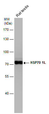
Rat tissue extract (50 µg) was separated by 7.5% SDS-PAGE, and the membrane was blotted with HSP70 1L antibody [N1N3] diluted at 1:1000. The HRP-conjugated anti-rabbit IgG antibody was used to detect the primary antibody.Application
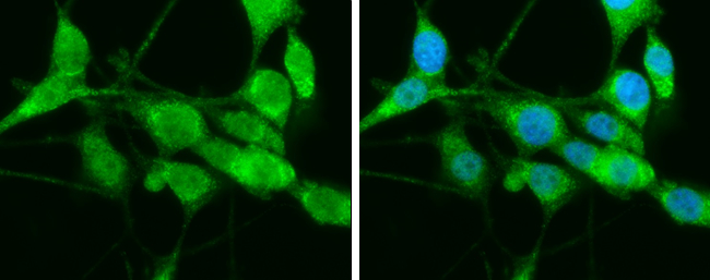
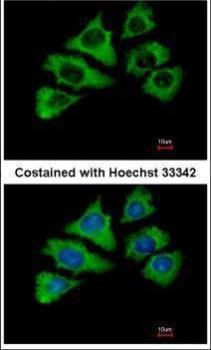
HSP70 1L antibody [N1N3] detects HSP70 1L protein at cytoplasm by immunofluorescent analysis. Sample: U87-MG cells were fixed in 4% paraformaldehyde at RT for 15 min. Green: HSP70 1L protein stained by HSP70 1L antibody [N1N3] diluted at 1:400. Blue: Hoechst 33342 staining.Application
Immunofluorescence analysis of methanol-fixed A549, using HSP70 1L antibody at 1:500 dilution.Application
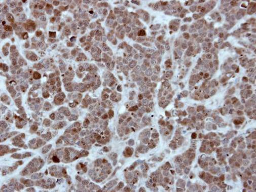
Immunohistochemical analysis of paraffin-embedded H661 xenograft, using HSP70 1L antibody at 1:500 dilution.Application
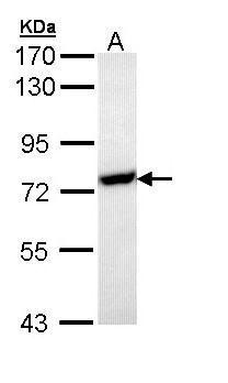
Sample (30 µg of whole cell lysate) A: Molt-4 7.5% SDS PAGE diluted at 1:1000 The HRP-conjugated anti-rabbit IgG antibody was used to detect the primary antibody.| Product Name | HSP70 1L antibody |
|---|---|
| Antibody Type | Primary Antibodies |
| Product description | Rabbit Polyclonal antibody to HSP70 1L (heat shock 70kDa protein 1-like)This gene encodes a 70kDa heat shock protein. In conjunction with other heat shock proteins, this protein stabilizes existing proteins against aggregation and mediates the folding of newly translated proteins in the cytosol and in organelles. The gene is located in the major histocompatibility complex class III region, in a cluster with two closely related genes which also encode isoforms of the 70kDa heat shock protein. [provided by RefSeq] |
| Immunogen | Recombinant protein encompassing a sequence within the center region of human HSP70 1L. The exact sequence is proprietary. |
| Clonality | Polyclonal |
|---|---|
| Isotype | IgG |
| Host Species | Rabbit |
| Tested Applications | ICC/IFIHC-PWB |
| *Optimal dilutions/concentrations should be determined by the researcher.
: |
|
| Species Reactivity | HumanMouseRat |
| Predicted Reactivity | Xenopus laevis, Pig, BovineXenopus laevis (81%), Pig (96%), Bovine (95%) |
| Concentration | 1mg/ml (Please refer to the vial label for the specific concentration) |
| Purification | Affinity purified |
| Gene Synonyms | HSP70-1L antibody HSP70-HOM antibody HSP70T antibody hum70t antibody HSPA1L antibody heat shock 10kDa protein 1-like antibody heat shock 70 kDa protein 1L antibody heat shock 70kD protein-like 1 antibody heat shock 70 kDa protein 1-Hom antibody he |
|---|---|
| Molecular Weight(MW) | 70 kDa (note) |
Application
Various whole cell extracts (30 µg) were separated by 7.5% SDS-PAGE, and the membrane was blotted with HSP70 1L antibody [N1N3] diluted at 1:1000. The HRP-conjugated anti-rabbit IgG antibody was used to detect the primary antibody.
Application
Mouse tissue extract (50 µg) was separated by 7.5% SDS-PAGE, and the membrane was blotted with HSP70 1L antibody [N1N3] diluted at 1:1000. The HRP-conjugated anti-rabbit IgG antibody was used to detect the primary antibody.
Application
Rat tissue extract (50 µg) was separated by 7.5% SDS-PAGE, and the membrane was blotted with HSP70 1L antibody [N1N3] diluted at 1:1000. The HRP-conjugated anti-rabbit IgG antibody was used to detect the primary antibody.
Application
HSP70 1L antibody [N1N3] detects HSP70 1L protein at cytoplasm by immunofluorescent analysis. Sample: U87-MG cells were fixed in 4% paraformaldehyde at RT for 15 min. Green: HSP70 1L protein stained by HSP70 1L antibody [N1N3] diluted at 1:400. Blue: Hoechst 33342 staining.
Application
Immunofluorescence analysis of methanol-fixed A549, using HSP70 1L antibody at 1:500 dilution.
Application
Immunohistochemical analysis of paraffin-embedded H661 xenograft, using HSP70 1L antibody at 1:500 dilution.
Application
Sample (30 µg of whole cell lysate) A: Molt-4 7.5% SDS PAGE diluted at 1:1000 The HRP-conjugated anti-rabbit IgG antibody was used to detect the primary antibody.| Positive Control | 293T , A431 , H1299 , HeLa , HepG2 , Molt-4 , Raji , mouse testis , rat testis , U87-MG , SK-N-SH , IMR32 , SK-N-AS |
|---|---|
| Application Notes | *Optimal dilutions/concentrations should be determined by the researcher.
: |
| Form | Liquid |
|---|---|
| Storage Instructions | Keep as concentrated solution. Aliquot and store at -20ºC or below. Avoid multiple freeze-thaw cycles. |
| Storage Buffer | 0.1M Tris, 0.1M Glycine, 10% Glycerol (pH7). 0.01% Thimerosal was added as a preservative. |


