Application
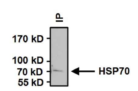
Immunoprecipitation of HSP70 in HeLa cells. Antigen-antibody complexes were formed by incubating 500μg whole cell lysate with 2μg of HSP70 monoclonal antibody overnight on a rocking platform at 4°C. The immune complexes were captured on 50μl Protein A/G Agarose , washed extensively, and eluted. Samples were then resolved on a 4-20% Tris-HCl polyacrylamide gel, transferred to a PVDF membrane, and blocked with 5% BSA/TBST for at least 1 hour. The membrane was probed with HSP70 antibody [5A5] at a dilution of 1:1000 overnight rotating at 4°C, washed in TBST, and probed with a proper secondary antibody. Chemiluminescent detection was performed.Application
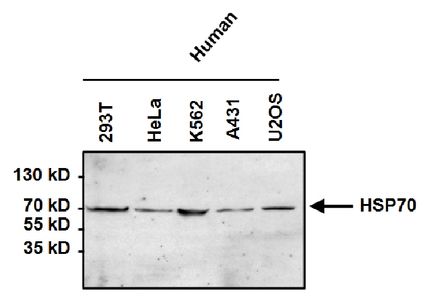
Western blot analysis of HSP70 in 50μg of various cell lysates. Proteins were transferred to a PVDF membrane and blocked with 5% BSA/TBST for at least 1 hour. The membrane was probed with HSP70 antibody [5A5] at a dilution of 1:1000 overnight at 4°C on a rocking platform, washed in TBS-0.1%Tween 20, and probed with a proper secondary antibody. Chemiluminescent detection was performed.Application
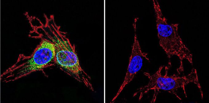
Immunofluorescent analysis of HSP70 in C6 glioma cells. HSP70 staining (green), F-Actin staining with Phalloidin (red) and nuclei with DAPI (blue) is shown. Cells were grown on slides and fixed with formaldehyde prior to staining. Cells were probed without (control) or with HSP70 antibody [5A5] at a dilution of 1:100-1:200 over night at 4 °C, washed with PBS and incubated with a proper secondary antibody. Imges were taken at 60X magnification.| Product Name | HSP70 antibody |
|---|---|
| Antibody Type | Primary Antibodies |
| Product description | Mouse monoclonal antibody [5A5] to HSP70This intronless gene encodes a 70kDa heat shock protein which is a member of the heat shock protein 70 family. In conjuction with other heat shock proteins, this protein stabilizes existing proteins against aggregation and mediates the folding of newly translated proteins in the cytosol and in organelles. It is also involved in the ubiquitin-proteasome pathway through interaction with the AU-rich element RNA-binding protein 1. The gene is located in the major histocompatibility complex class III region, in a cluster with two closely related genes which encode similar proteins. [provided by RefSeq, Jul 2008] |
| Immunogen | Human recombinant HSP70 over expressed in E. coli. |
| Clonality | Monoclonal |
|---|---|
| Isotype | IgG1 |
| Clone Number | 5A5 |
| Host Species | Mouse |
| Tested Applications | BlockingFACSGSAICC/IFIHCIHC-FIPWB |
| ICC/IF: 1:50 - 1:3000: |
|
| Species Reactivity | HumanMouseDrosophilaRatYeastAmphibiansFishHamsterPrimateRabbitBovineAvian |
| Concentration | 1mg/ml |
| Gene Synonyms | HSPA1 HSP70-1A HSP70-1 heat shock 70kDa protein 1A HSP70I HSP72 HSPA1A |
|---|---|
| Target | HSP70/Heat Shock Protein 70 |
| Tissue Specificity | This antibody detects several members of HSP70 family including HSP70, HSC70, GRP78, and following heat shock, HSP72 from yeast, rat, Drosophila, fish, mouse, avian, amphibian and human samples. |
Application
Immunoprecipitation of HSP70 in HeLa cells. Antigen-antibody complexes were formed by incubating 500μg whole cell lysate with 2μg of HSP70 monoclonal antibody overnight on a rocking platform at 4°C. The immune complexes were captured on 50μl Protein A/G Agarose , washed extensively, and eluted. Samples were then resolved on a 4-20% Tris-HCl polyacrylamide gel, transferred to a PVDF membrane, and blocked with 5% BSA/TBST for at least 1 hour. The membrane was probed with HSP70 antibody [5A5] at a dilution of 1:1000 overnight rotating at 4°C, washed in TBST, and probed with a proper secondary antibody. Chemiluminescent detection was performed.
Application
Western blot analysis of HSP70 in 50μg of various cell lysates. Proteins were transferred to a PVDF membrane and blocked with 5% BSA/TBST for at least 1 hour. The membrane was probed with HSP70 antibody [5A5] at a dilution of 1:1000 overnight at 4°C on a rocking platform, washed in TBS-0.1%Tween 20, and probed with a proper secondary antibody. Chemiluminescent detection was performed.
Application
Immunofluorescent analysis of HSP70 in C6 glioma cells. HSP70 staining (green), F-Actin staining with Phalloidin (red) and nuclei with DAPI (blue) is shown. Cells were grown on slides and fixed with formaldehyde prior to staining. Cells were probed without (control) or with HSP70 antibody [5A5] at a dilution of 1:100-1:200 over night at 4 °C, washed with PBS and incubated with a proper secondary antibody. Imges were taken at 60X magnification.| Application Notes | ICC/IF: 1:50 - 1:3000: |
|---|
| Form | Liquid |
|---|---|
| Storage Instructions | -20° C, Avoid Freeze/Thaw Cycles |
| Storage Buffer | Ascites containing 0.05 % sodium azide |


