Application
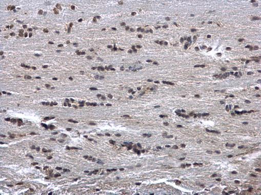
HUS1 antibody detects HUS1 protein at cytosol and nucleus on rat fore brain by immunohistochemical analysis. Sample: Paraffin-embedded rat fore brain. HUS1 antibody dilution: 1:500.Application
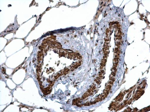
HUS1 antibody detects HUS1 protein at cytosol and nucleus on mouse lymph node by immunohistochemical analysis. Sample: Paraffin-embedded mouse lymph node. HUS1 antibody dilution: 1:500.Application
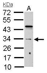
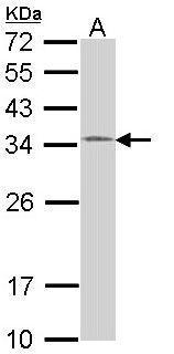
HUS1 antibody detects HUS1 protein by Western blot analysis. A. 30 µg C8D30 whole cell lysate/extract 12 % SDS-PAGE HUS1 antibody dilution: 1:1000Application
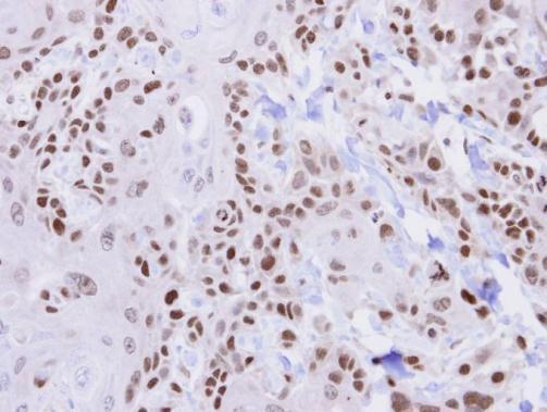
Immunohistochemical analysis of paraffin-embedded CA922 xenograft , using HUS1 antibody at 1:500 dilution.Application
Sample (30 ug of whole cell lysate) A: 293T 12% SDS PAGE diluted at 1:1000| Product Name | HUS1 antibody |
|---|---|
| Antibody Type | Primary Antibodies |
| Product description | Rabbit Polyclonal antibody to HUS1 (HUS1 checkpoint homolog (S. pombe))The protein encoded by this gene is a component of an evolutionarily conserved, genotoxin-activated checkpoint complex that is involved in the cell cycle arrest in response to DNA damage. This protein forms a heterotrimeric complex with checkpoint proteins RAD9 and RAD1. In response to DNA damage, the trimeric complex interacts with another protein complex consisting of checkpoint protein RAD17 and four small subunits of the replication factor C (RFC), which loads the combined complex onto the chromatin. The DNA damage induced chromatin binding has been shown to depend on the activation of the checkpoint kinase ATM, and is thought to be an early checkpoint signaling event. [provided by RefSeq] |
| Immunogen | Recombinant protein encompassing a sequence within the center region of human HUS1. The exact sequence is proprietary. |
| Clonality | Polyclonal |
|---|---|
| Isotype | IgG |
| Host Species | Rabbit |
| Tested Applications | IHC-PWB |
| *Optimal dilutions/concentrations should be determined by the researcher.
: |
|
| Species Reactivity | HumanMouseRat |
| Predicted Reactivity | PigPig (89%) |
| Concentration | 1mg/ml |
| Purification | Affinity purified |
| Gene Synonyms | hHUS1 antibody HUS1 antibody checkpoint protein HUS1 antibody hus1+-like protein antibody HUS1 checkpoint homolog (S. pombe) antibody |
|---|---|
| Molecular Weight(MW) | 32 kDa (note) (2) |
| Cellular Localization | Nucleus , Cytoplasm |
Application
HUS1 antibody detects HUS1 protein at cytosol and nucleus on rat fore brain by immunohistochemical analysis. Sample: Paraffin-embedded rat fore brain. HUS1 antibody dilution: 1:500.
Application
HUS1 antibody detects HUS1 protein at cytosol and nucleus on mouse lymph node by immunohistochemical analysis. Sample: Paraffin-embedded mouse lymph node. HUS1 antibody dilution: 1:500.
Application
HUS1 antibody detects HUS1 protein by Western blot analysis. A. 30 µg C8D30 whole cell lysate/extract 12 % SDS-PAGE HUS1 antibody dilution: 1:1000
Application
Immunohistochemical analysis of paraffin-embedded CA922 xenograft , using HUS1 antibody at 1:500 dilution.
Application
Sample (30 ug of whole cell lysate) A: 293T 12% SDS PAGE diluted at 1:1000| Positive Control | 293T , C8D30 |
|---|---|
| Application Notes | *Optimal dilutions/concentrations should be determined by the researcher.
: |
| Form | Liquid |
|---|---|
| Storage Instructions | Keep as concentrated solution. Aliquot and store at -20ºC or below. Avoid multiple freeze-thaw cycles. |
| Storage Buffer | 0.1M Tris, 0.1M Glycine, 20% Glycerol (pH7). 0.01% Thimerosal was added as a preservative. |


