Application
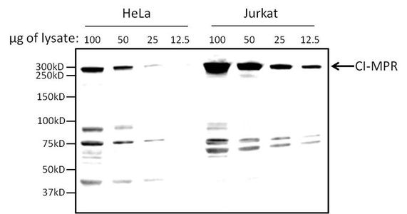
WB analysis analysis of various amounts of HeLa and Jurkat lysates using CI-MPR antibody at a dilution of 1:2,000.Application
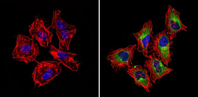
ICC/IF analysis of HeLa cells with (right) or without (left) CI-MPR antibody (green) at a dilution of 1:100. F-actin (red) was stained with a fluorescent red phalloidin and nuclei (blue) were stained with DAPI. Images were taken at a magnification of 60x.Application
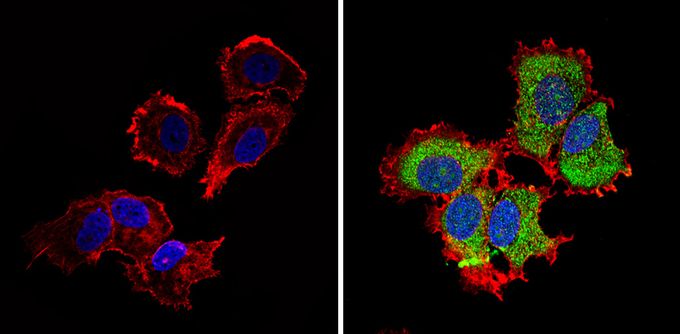
ICC/IF analysis of MCF-7 cells with (right) or without (left) CI-MPR antibody (green) at a dilution of 1:100. F-actin (red) was stained with a fluorescent red phalloidin and nuclei (blue) were stained with DAPI. Images were taken at a magnification of 60x.Application
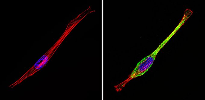
ICC/IF analysis of NIH-3T3 cells with (right) or without (left) CI-MPR antibody at a dilution of 1:100. F-actin (red) was stained with a fluorescent red phalloidin and nuclei (blue) were stained with DAPI. Images were taken at a magnification of 60x.| Product Name | IGF2 receptor antibody |
|---|---|
| Antibody Type | Primary Antibodies |
| Product description | Rabbit Polyclonal antibody to IGF2 receptorThe cation-independent mannose-6-phosphate receptor (CI-MPR) is a multifunctional, type I integral membrane glycoprotein, acting as both an intracellular protein carrier and a cell surface signaling molecule. Functionally, CI-MPR binds newly synthesized mannose-6-phosphate and transports it to the lysosome in mammalian cells. |
| Immunogen | 300 kDa cation-independent receptor purified from adult bovine liver tissue. |
| Clonality | Polyclonal |
|---|---|
| Host Species | Rabbit |
| Tested Applications | DotICC/IFWB |
| ICC/IF: 1:50 - 1:500 | |
| Species Reactivity | HumanMousePrimateBovine |
| Concentration | 1mg/ml |
| Purification | Affinity purified |
| Target | IGF2 receptor |
|---|
Application
WB analysis analysis of various amounts of HeLa and Jurkat lysates using CI-MPR antibody at a dilution of 1:2,000.
Application
ICC/IF analysis of HeLa cells with (right) or without (left) CI-MPR antibody (green) at a dilution of 1:100. F-actin (red) was stained with a fluorescent red phalloidin and nuclei (blue) were stained with DAPI. Images were taken at a magnification of 60x.
Application
ICC/IF analysis of MCF-7 cells with (right) or without (left) CI-MPR antibody (green) at a dilution of 1:100. F-actin (red) was stained with a fluorescent red phalloidin and nuclei (blue) were stained with DAPI. Images were taken at a magnification of 60x.
Application
ICC/IF analysis of NIH-3T3 cells with (right) or without (left) CI-MPR antibody at a dilution of 1:100. F-actin (red) was stained with a fluorescent red phalloidin and nuclei (blue) were stained with DAPI. Images were taken at a magnification of 60x.| Application Notes | ICC/IF: 1:50 - 1:500 |
|---|
| Form | Liquid |
|---|---|
| Storage Instructions | Keep as concentrated solution. Aliquot and store at -20ºC or below. Avoid multiple freeze-thaw cycles. |
| Storage Buffer | Antisera containing 0.05% sodium azide. |


