Application
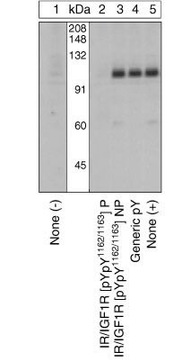
Extracts of CHO-T cells over-expressing the human insulin receptor unstimulated (1) or stimulated with 100 nM insulin for 10 min at 37°C (2-5) were resolved by SDS-PAGE on a 10% Tris-glycine gel and transferred to PVDF. The membrane was blocked with a 5% BSA-TBST buffer overnight at 4°C and incubated with the IR/IGF1R [pYpY1162/1163] antibody for two hours at room temperature in a 3% BSA-TBST buffer, following prior incubation with: no peptide (1, 5), the phosphopeptide immunogen (2), the non-phosphorylated peptide corresponding to the phosphopeptide immunogen (3), or a generic phosphotyrosine-containing peptide (4). After washing, the membrane was incubated with goat F(ab)2 anti-rabbit IgG HRP-conjugate and signals were detected. The data show that only the phosphopeptide corresponding to IR/IGF1R [pYpY1162/1163] completely blocks the antibody signal, demonstrating the specificity of the antibody. The data also show the up-regulation of this site upon stimulation with insulin in this cell system.| Product Name | Insulin receptor (phospho Tyr1162/Tyr1163) antibody |
|---|---|
| Antibody Type | Primary Antibodies |
| Product description | Rabbit polyclonal to Insulin Receptor / IGF1R ( phospho Y1162 + Y1163 )Biological actions of Insulin and IGF1 are mediated by their respective cell surface receptor tyrosine kinases that regulate multiple signaling pathways through activation of a series of phosphorylation cascades. The Insulin Receptor and IGF1R are heterotetrameric proteins consisting of two ligand-binding alpha subunits and two beta subunits that each contain a tyrosine kinase domain. Insulin/IGF1 binding to the extracellular domain leads to auto-phosphorylation of the receptor and activation of the intrinsic tyrosine kinase activity, leading to tyrosine phosphorylation of downstream target proteins. These two receptors differ in sequence in regions that confer specificity for the designated ligand as well as in certain intracellular signaling domains, resulting in significant differences in the functional consequences of activation of each receptor. The catalytic loops within the tyrosine kinase domains of the Insulin Receptor/IGF1R contain a three tyrosine motif corresponding to tyrosines 1158, 1162 and 1163 (for the IR) and 1131, 1135 and 1136 (for the IGF1R). It is generally believed that autophosphorylation within the activation loop proceeds in a processive manner initiating at the second tyrosine (1162 or 1135), followed by phosphorylation at the first tyrosine (1158 or 1131), then the last (1163 or 1136), upon which the Insulin Receptor or IGF1R becomes fully active. |
| Immunogen | Synthetic phosphopeptide derived from the region of Insulin Receptor/IGF1R that contains tyrosines 1162 and 1163. The corresponding residues in the IGF1R are 1135 and 1136. |
| Modification | p-Tyr1162/Tyr1163 |
| Clonality | Polyclonal |
|---|---|
| Isotype | IgG |
| Host Species | Rabbit |
| Tested Applications | WB |
| WB: Use at a dilution of 1/1000. Detects a band of approximately 120 kDa. Optimal dilutions/concentrations should be determined by the end user.: |
|
| Species Reactivity | HumanMouseRat |
| Concentration | 1mg/ml |
| Purification | Affinity purified |
| Gene Synonyms | Insulin receptor (phospho Y1162/Y116) Insulin receptor (phospho Tyr1162/Tyr116) Insulin receptor (pY1162/pY116) Insulin receptor (pTyr1162/pTyr116) Insulin receptor phospho Y1162/Y116 Insulin receptor phospho Tyr1162/Tyr116 Phospho Insulin receptor |
|---|---|
| Target | Insulin receptor |
| Tissue Specificity | Although exhibiting a preference for IR/IGF-1R, this antibody has been shown by both peptide competition and protein blotting to react with other dual phosphotyrosine motifs from proteins such as c-Met and Shc. |
| Cellular Localization | Type I membrane protein |
Application
Extracts of CHO-T cells over-expressing the human insulin receptor unstimulated (1) or stimulated with 100 nM insulin for 10 min at 37°C (2-5) were resolved by SDS-PAGE on a 10% Tris-glycine gel and transferred to PVDF. The membrane was blocked with a 5% BSA-TBST buffer overnight at 4°C and incubated with the IR/IGF1R [pYpY1162/1163] antibody for two hours at room temperature in a 3% BSA-TBST buffer, following prior incubation with: no peptide (1, 5), the phosphopeptide immunogen (2), the non-phosphorylated peptide corresponding to the phosphopeptide immunogen (3), or a generic phosphotyrosine-containing peptide (4). After washing, the membrane was incubated with goat F(ab)2 anti-rabbit IgG HRP-conjugate and signals were detected. The data show that only the phosphopeptide corresponding to IR/IGF1R [pYpY1162/1163] completely blocks the antibody signal, demonstrating the specificity of the antibody. The data also show the up-regulation of this site upon stimulation with insulin in this cell system.| Positive Control | CHO-T cells transfected with a vector containing insulin receptor and stimulated with 100 nM insulin for 10 minutes at 37ºC. |
|---|---|
| Application Notes | WB: Use at a dilution of 1/1000. Detects a band of approximately 120 kDa. Optimal dilutions/concentrations should be determined by the end user.: |
| Form | Liquid |
|---|---|
| Storage Instructions | Stable at 2 years at -20 degrees Celcius from the date of shipment. |
| Storage Buffer | Preservative: 0.05% Sodium Azide; Constituents: 50% Glycerol, PBS, 1mg/ml BSA. pH 7.3 |


