Application
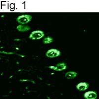
IHC analysis of rat cerebellum using IP3 Receptor I antibody.| Product Name | IP3 Receptor I antibody |
|---|---|
| Antibody Type | Primary Antibodies |
| Product description | Rabbit Polyclonal antibody to IP3 Receptor IInositol 1,4,5-trisphosphate (IP3) is a second messenger for many growth factors, hormones, and neurotransmitters. Upon binding to the IP3 receptor (IP3R), IP3 triggers the release of intracellular, luminal calcium to the cytosol. Functional IP3R is a homo- or heterotetramer of ~240 kDa glycoprotein subunits, that is structurally reminiscent of members of the voltage-gated ion channel superfamily. IP3R protein is structurally and functionally related to one other important intracellular calcium-release channel, the ryanodine receptor. The similarity between the two receptors continues at the physiological level owing to a physical association each receptor can have with the immunophilin protein, FKBP12. Cytosolic calcium levels appear to be regulated by a feedback loop that starts with calcium activation of protein kinase C (PKC) and calcineurin, a protein phosphatase. Phosphorylation of the IP3R by PKC causes an increase in IP3-mediated calcium release. Concomitantly, the phosphatase activity of calcineurin is stimulated upon its association with the FKBP12-IP3R complex. Calcium release is reduced when the PKC target site on the IP3R is dephosphorylated by calcineurin resulting in calcium oscillations. Mammalian IP3R subunits are the product of three distinct genes that are widely expressed and differentially regulated. IP3R type I (IP3R-I) has been detected in heart, liver, kidney, ovary, and Purkinje neurons of the cerebellum. IP3R-II is found predominantly in the brain. IP3R-III is known to be expressed in pancreatic islets, kidney, and the gastrointestinal tract. |
| Immunogen | Synthetic Peptide: N(1829) K K K D D E V D R D A P S R K K A K E(1848) |
| Clonality | Polyclonal |
|---|---|
| Host Species | Rabbit |
| Tested Applications | FACSICC/IFIHC-FIPWB |
|
For WB: Use at a dilution of 1:1000.: |
|
| Species Reactivity | HumanMouseDogRatHamster |
| Concentration | 1mg/ml |
| Purification | Affinity purified |
| Gene Synonyms | Itpr1 I145TR InsP3R InsP3R1 inositol 1 4 5-triphosphate receptor type 1 |
|---|---|
| Target | IP3 Receptor I |
Application
IHC analysis of rat cerebellum using IP3 Receptor I antibody.| Application Notes |
For WB: Use at a dilution of 1:1000.: |
|---|
| Form | Liquid |
|---|---|
| Storage Instructions | Keep as concentrated solution. Aliquot and store at -20ºC or below. Avoid multiple freeze-thaw cycles. |
| Storage Buffer | PBS containing 1 mg/ml BSA and 0.05% sodium azide. |


