Application
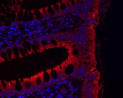
Immunohistochemical analysis of paraffin-embedded human colon, using JAM-B antibody at 1:200 dilution.(Image courtesy of Sergei Grivennikov Ph.D (Laboratory of Dr. Michael Karin, UCSD.)Application
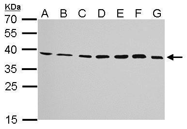
JAM-B antibody detects JAM2 protein by western blot analysis. A. 30 µg Neuro2A whole cell lysate/extract B. 30 µg GL261 whole cell lysate/extract C. 30 µg C8D30 whole cell lysate/extract D. 30 µg NIH-3T3 whole cell lysate/extract E. 30 µg BCL-1 whole cell lysate/extract F. 30 µg Raw264.7 whole cell lysate/extract G. 30 µg C2C12 whole cell lysate/extract 12% SDS-PAGE JAM-B antibody dilution: 1:1000 The HRP-conjugated anti-rabbit IgG antibody was used to detect the primary antibody.Application
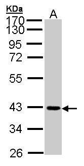
Sample (30 µg of whole cell lysate) A: A431 12% SDS PAGE diluted at 1:1000 The HRP-conjugated anti-rabbit IgG antibody was used to detect the primary antibody.Application
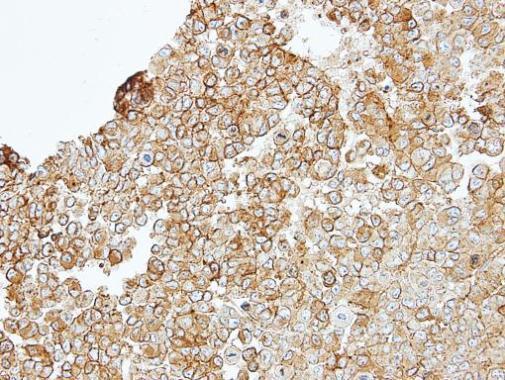
Immunohistochemical analysis of paraffin-embedded TOV-112D xenograft, using JAM-B antibody at 1:100 dilution.Application
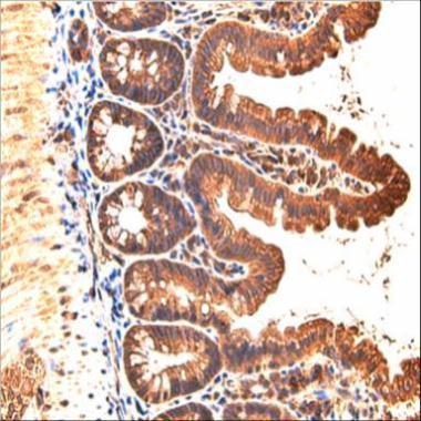
Immunohistochemical analysis of paraffin-embedded mouse small intestine, using JAM-B antibody at 1:200 dilution.(Image courtesy of Koji Taniguchi, Ph.D (Laboratory of Dr. Michael Karin, UCSD.)| Product Name | JAM-B antibody |
|---|---|
| Antibody Type | Primary Antibodies |
| Product description | Rabbit Polyclonal antibody to JAM-B (junctional adhesion molecule 2)Tight junctions represent one mode of cell-to-cell adhesion in epithelial or endothelial cell sheets, forming continuous seals around cells and serving as a physical barrier to prevent solutes and water from passing freely through the paracellular space. The protein encoded by this immunoglobulin superfamily gene member is localized in the tight junctions between high endothelial cells. It acts as an adhesive ligand for interacting with a variety of immune cell types and may play a role in lymphocyte homing to secondary lymphoid organs. [provided by RefSeq] |
| Immunogen | Recombinant protein encompassing a sequence within the center region of human JAM-B. The exact sequence is proprietary. |
| Clonality | Polyclonal |
|---|---|
| Isotype | IgG |
| Host Species | Rabbit |
| Tested Applications | IHCIHC-PWB |
| *Optimal dilutions/concentrations should be determined by the researcher.
: |
|
| Species Reactivity | HumanMouse |
| Predicted Reactivity | Rat, BovineRat (81%), Bovine (90%) |
| Concentration | 1mg/ml (Please refer to the vial label for the specific concentration) |
| Purification | Affinity purified |
| Gene Synonyms | C21orf43 antibody CD322 antibody JAM-B antibody JAMB antibody PRO245 antibody VE-JAM antibody VEJAM antibody JAM2 antibody JAM-IT/VE-JAM antibody JAM-2 antibody junctional adhesion molecule B antibody vascular endothelial junction-associated mol |
|---|---|
| Molecular Weight(MW) | 33 kDa (note) (2) |
| Cellular Localization | Cell junction , tight junction (By similarity) , Cell membrane; Single-pass type I membrane protein (By similarity) |
Application
Immunohistochemical analysis of paraffin-embedded human colon, using JAM-B antibody at 1:200 dilution.(Image courtesy of Sergei Grivennikov Ph.D (Laboratory of Dr. Michael Karin, UCSD.)
Application
JAM-B antibody detects JAM2 protein by western blot analysis. A. 30 µg Neuro2A whole cell lysate/extract B. 30 µg GL261 whole cell lysate/extract C. 30 µg C8D30 whole cell lysate/extract D. 30 µg NIH-3T3 whole cell lysate/extract E. 30 µg BCL-1 whole cell lysate/extract F. 30 µg Raw264.7 whole cell lysate/extract G. 30 µg C2C12 whole cell lysate/extract 12% SDS-PAGE JAM-B antibody dilution: 1:1000 The HRP-conjugated anti-rabbit IgG antibody was used to detect the primary antibody.
Application
Sample (30 µg of whole cell lysate) A: A431 12% SDS PAGE diluted at 1:1000 The HRP-conjugated anti-rabbit IgG antibody was used to detect the primary antibody.
Application
Immunohistochemical analysis of paraffin-embedded TOV-112D xenograft, using JAM-B antibody at 1:100 dilution.
Application
Immunohistochemical analysis of paraffin-embedded mouse small intestine, using JAM-B antibody at 1:200 dilution.(Image courtesy of Koji Taniguchi, Ph.D (Laboratory of Dr. Michael Karin, UCSD.)| Positive Control | A431 , Neuro2A , GL261 , C8D30 , NIH-3T3 , BCL-1 , Raw264.7 , C2C12 |
|---|---|
| Application Notes | *Optimal dilutions/concentrations should be determined by the researcher.
: |
| Form | Liquid |
|---|---|
| Storage Instructions | Keep as concentrated solution. Aliquot and store at -20ºC or below. Avoid multiple freeze-thaw cycles. |
| Storage Buffer | 0.1M Tris, 0.1M Glycine, 10% Glycerol (pH7). 0.01% Thimerosal was added as a preservative. |


