Application
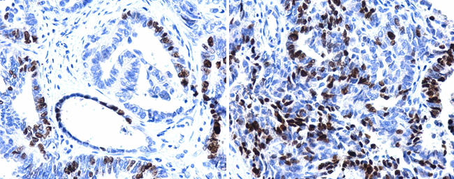
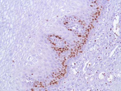
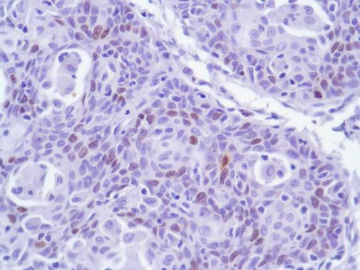
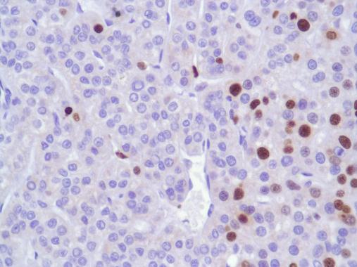
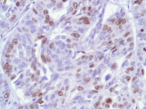
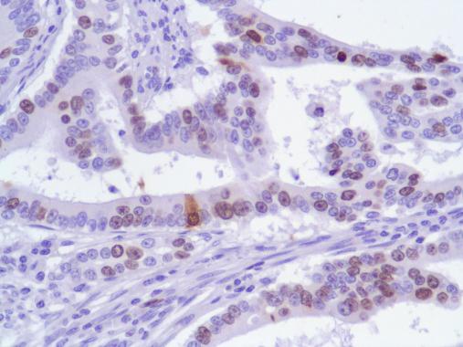
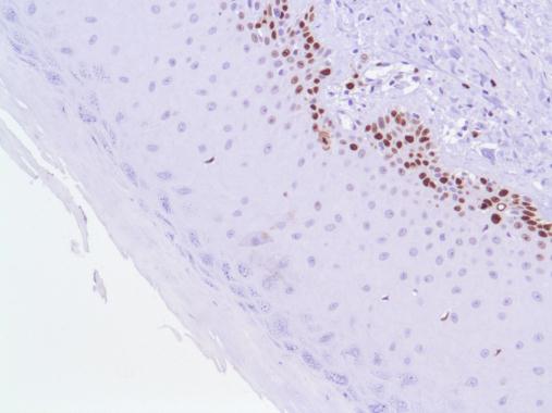
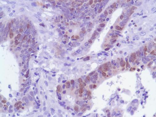
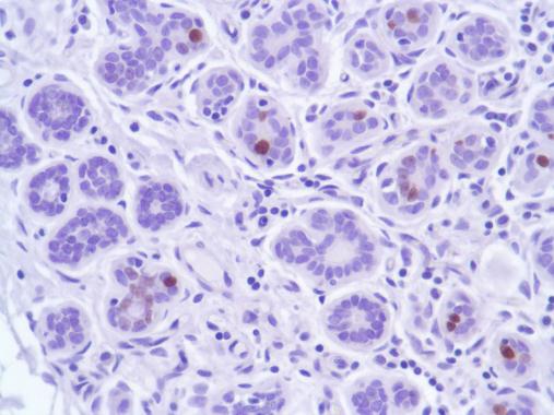
Ki67 antibody [SP6] detects Ki67 protein at nucleus in human cervical metaplasia (left) and carcinoma (right) by immunohistochemical analysis. Sample: Paraffin-embedded human cervix. Ki67 antibody [SP6] diluted at 1:200.Application
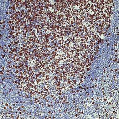
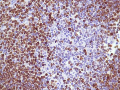
Formalin fixed paraffin embedded human tonsil stained with Ki-67 antibodyApplication
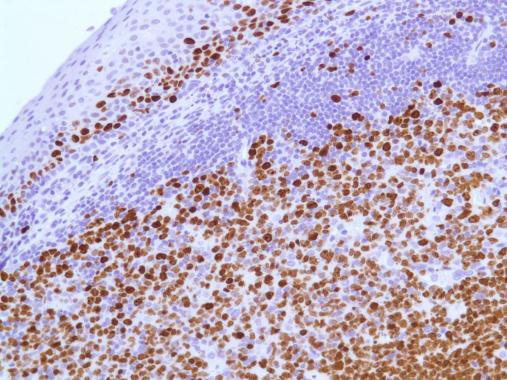
Human Tonsil stained with anti-Ki-67 antibodyApplication
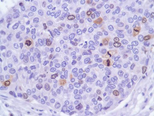
Human Bladder Transitional Cell Carcinoma stained with anti-Ki-67 antibodyApplication
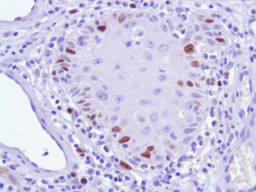
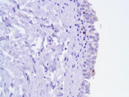
Human Esophagus stained with anti-Ki-67 antibodyApplication
Human Fallopian Tube stained with anti-Ki-67 antibodyApplication
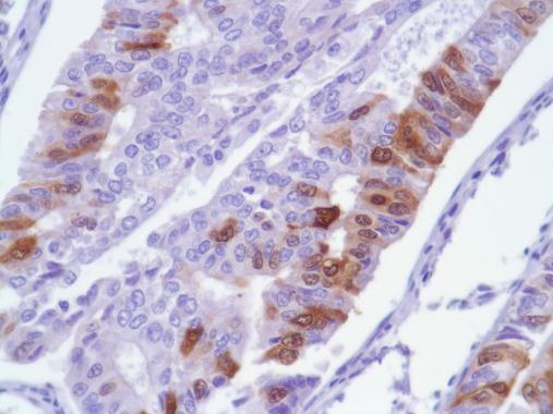
Human Endometrial Adenocarcinoma stained with anti-Ki-67 antibodyApplication
Human Cervical Squamous Cell Carcinoma stained with anti-Ki-67 antibodyApplication
Human Skin Squamous Cell Carcinoma stained with anti-Ki-67 antibodyApplication
Human Hepatocellular Carcinoma stained with anti-Ki-67 antibodyApplication
Human Lung Squamous Cell Carcinoma stained with anti-Ki-67 antibodyApplication
Human Stomach Adenocarcinoma stained with anti-Ki-67 antibodyApplication
Human Skin stained with anti-Ki-67 antibodyApplication
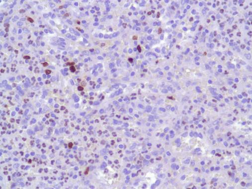
Human Thymus stained with anti-Ki-67 antibodyApplication
Human Spleen stained with anti-Ki-67 antibodyApplication
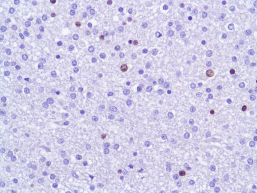
Human Spinal Cord stained with anti-Ki-67 antibodyApplication
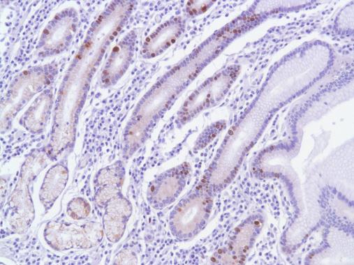
Human Stomach stained with anti-Ki-67 antibodyApplication
Immunohistochemical staining using Ki-67 antibodyApplication
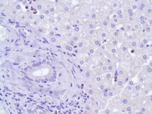
Human Liver stained with anti-Ki-67 antibodyApplication
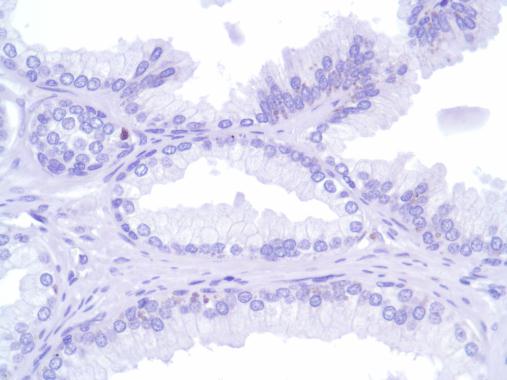
Human Prostate stained with anti-Ki-67 antibodyApplication
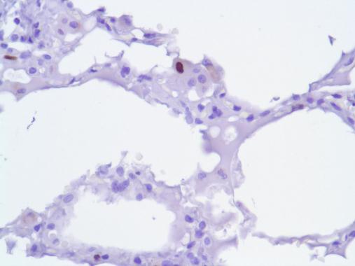
Human Lung stained with anti-Ki-67 antibodyApplication
Human Lung Adenocarcinoma stained with anti-Ki-67 antibodyApplication
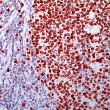
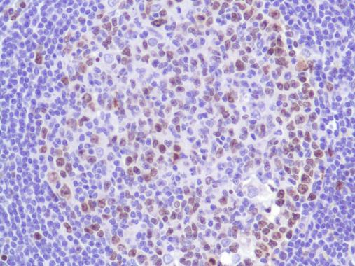
Human Lymph Node stained with anti-Ki-67 antibodyApplication
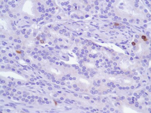
Human Prostate Adenocarcinoma stained with anti-Ki-67 antibodyApplication
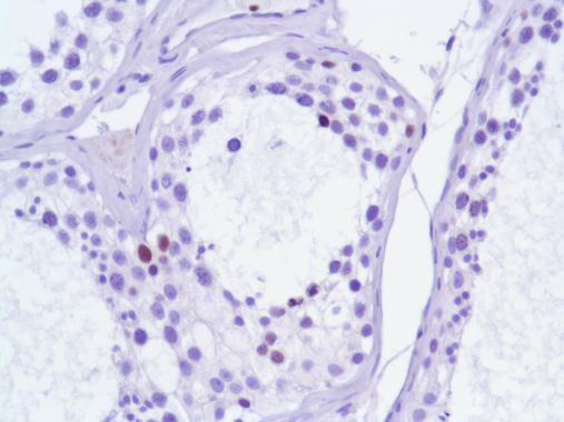
Human Testis stained with anti-Ki-67 antibodyApplication
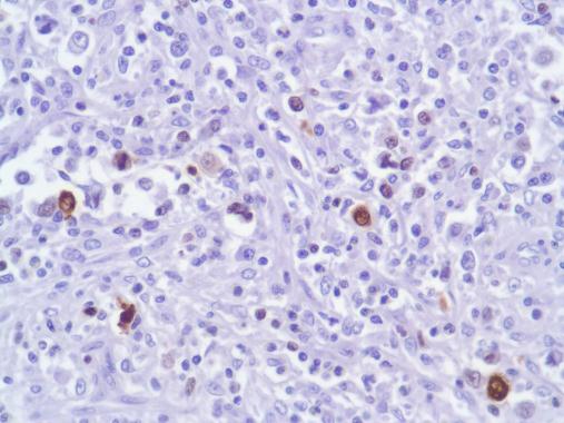
Human B Cell Lymphoma stained with anti-Ki-67 antibodyApplication
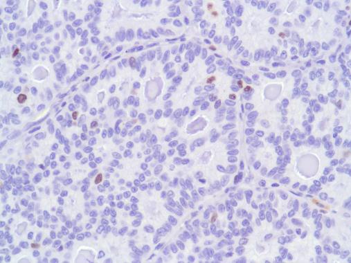
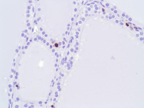
Human Thyroid stained with anti-Ki-67 antibodyApplication
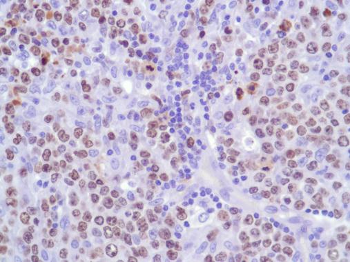
Human HK Lymphoma stained with anti-Ki-67 antibodyApplication
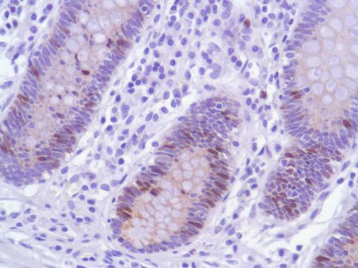
Human Colon stained with anti-Ki-67 antibodyApplication
Human Colon Adenocarcinoma stained with anti-Ki-67 antibodyApplication
Human Breast stained with anti-Ki-67 antibodyApplication
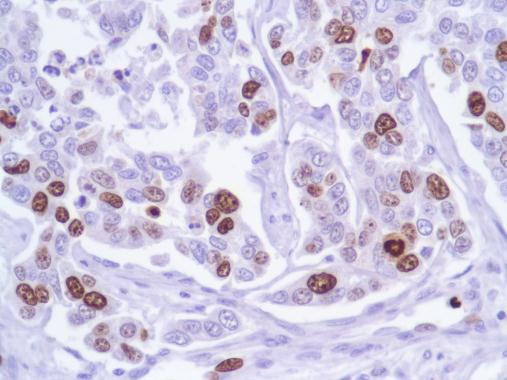
Human Breast Adenocarcinoma stained with anti-Ki-67 antibody| Product Name | Ki67 antibody [SP6] |
|---|---|
| Antibody Type | Primary Antibodies |
| Product description | Rabbit monoclonal [SP6] to Ki67 - Proliferation MarkerKi67 antigen is the prototypic cell cycle related nuclear protein, expressed by proliferating cells in all phases of the active cell cycle (G1, S, G2 and M phase). It is absent in resting (G0) cells. Ki67 antibodies are useful in establishing the cell growing fraction in neoplasms (immunohistochemically quantified by determining the number of Ki67-positive cells among the total number of resting cells = Ki67 index). In neoplastic tissues the prognostic value is comparable to the tritiated thymidine-labelling index. The correlation between low Ki67 index and histologically low-grade tumours is strong. |
| Clonality | Monoclonal |
|---|---|
| Isotype | IgG |
| Clone Number | SP6 |
| Host Species | Rabbit |
| Tested Applications | ICC/IF,IHC,IHC-F,IHC-P,WB |
| IHC-P: 1:100-1:1000. Staining of formalin-fixed tissues is required by boiling tissue sections in 10mM citrate buffer, pH 6.0 for 10 min followed by cooling at RT for 20 min. WB: 1/200. Predicted molecular weight: 359 kDa. Not tested in other applications | |
| Species Reactivity | Human,Mouse,Rat,Zebra Fish |
| Concentration | 1mg/ml |
| Purification | Unpurified |
| Gene Synonyms | KIA antibody, Proliferation related Ki67 antigen antibody, Proliferation related Ki 67 antigen antibody, Antigen KI-67 antibody, Antigen KI67 antibody, Antigen identified by monoclonal antibody Ki67 antibody, Antigen identified by monoclonal antibody |
|---|---|
| Target | Ki67 |
| Cellular Localization | Nuclear |
Application
Ki67 antibody [SP6] detects Ki67 protein at nucleus in human cervical metaplasia (left) and carcinoma (right) by immunohistochemical analysis. Sample: Paraffin-embedded human cervix. Ki67 antibody [SP6] diluted at 1:200.
Application
Formalin fixed paraffin embedded human tonsil stained with Ki-67 antibody
Application
Human Tonsil stained with anti-Ki-67 antibody
Application
Human Bladder Transitional Cell Carcinoma stained with anti-Ki-67 antibody
Application
Human Esophagus stained with anti-Ki-67 antibody
Application
Human Fallopian Tube stained with anti-Ki-67 antibody
Application
Human Endometrial Adenocarcinoma stained with anti-Ki-67 antibody
Application
Human Cervical Squamous Cell Carcinoma stained with anti-Ki-67 antibody
Application
Human Skin Squamous Cell Carcinoma stained with anti-Ki-67 antibody
Application
Human Hepatocellular Carcinoma stained with anti-Ki-67 antibody
Application
Human Lung Squamous Cell Carcinoma stained with anti-Ki-67 antibody
Application
Human Stomach Adenocarcinoma stained with anti-Ki-67 antibody
Application
Human Skin stained with anti-Ki-67 antibody
Application
Human Thymus stained with anti-Ki-67 antibody
Application
Human Spleen stained with anti-Ki-67 antibody
Application
Human Spinal Cord stained with anti-Ki-67 antibody
Application
Human Stomach stained with anti-Ki-67 antibody
Application
Immunohistochemical staining using Ki-67 antibody
Application
Human Liver stained with anti-Ki-67 antibody
Application
Human Prostate stained with anti-Ki-67 antibody
Application
Human Lung stained with anti-Ki-67 antibody
Application
Human Lung Adenocarcinoma stained with anti-Ki-67 antibody
Application
Human Lymph Node stained with anti-Ki-67 antibody
Application
Human Prostate Adenocarcinoma stained with anti-Ki-67 antibody
Application
Human Testis stained with anti-Ki-67 antibody
Application
Human B Cell Lymphoma stained with anti-Ki-67 antibody
Application
Human Thyroid stained with anti-Ki-67 antibody
Application
Human HK Lymphoma stained with anti-Ki-67 antibody
Application
Human Colon stained with anti-Ki-67 antibody
Application
Human Colon Adenocarcinoma stained with anti-Ki-67 antibody
Application
Human Breast stained with anti-Ki-67 antibody
Application
Human Breast Adenocarcinoma stained with anti-Ki-67 antibody| Application Notes | IHC-P: 1:100-1:1000. Staining of formalin-fixed tissues is required by boiling tissue sections in 10mM citrate buffer, pH 6.0 for 10 min followed by cooling at RT for 20 min. WB: 1/200. Predicted molecular weight: 359 kDa. Not tested in other applications |
|---|
| Form | Liquid |
|---|---|
| Storage Instructions | Keep as concentrated solution, aliquot and store at 4°C. Do not freeze. |
| Storage Buffer | Phosphate-buffered saline, pH 7.4 containing 1% BSA and 0.09% sodium azide |


