Application
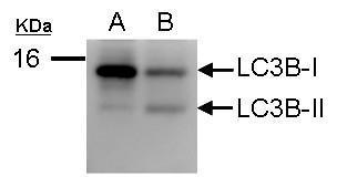
Sample (50 ug of whole cell lysate) A: Huh7 B: virus infected Huh7 12% SDS PAGE diluted at 1:1000Application
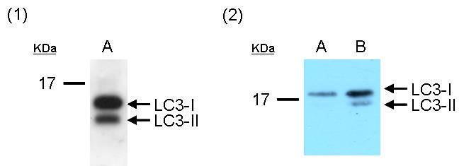
Western blot analysis of LC3 protein in PC-3 cell line. (1) 20ug of whole cell lysate was separated by using Urea gel (6M Urea in 13.5% SDS gel) and detected with unpurified serum diluted at 1:4000. (2) PC-3 cells were cultured with serum-containing medium (A) and serum-free medium (B) for 3 days. 5ug of whole cell lysate was separated by using Urea gel (6M Urea in 13.5% SDS gel) and detected with antibody diluted at 1:5000.Application
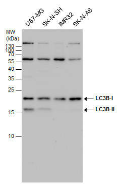
LC3B antibody detects LC3B protein by western blot analysis. Various whole cell extracts (30 μg) were separated by 15 % SDS-PAGE, and blotted with LC3B antibody diluted by 1:500Application
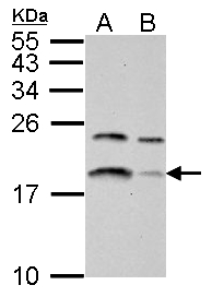
LC3B antibody detects LC3B protein by western blot analysis. A. 30 μg U87-MG whole cell lysate/extract B. 30 μg SK-N-SH whole cell lysate/extract 15 % SDS-PAGE LC3B antibody dilution: 1:500Application
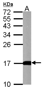
LC3B antibody detects LC3B protein by western blot analysis. A. 50 μg mouse brain lysate/extract 15 % SDS-PAGE LC3B antibody dilution: 1:500Application
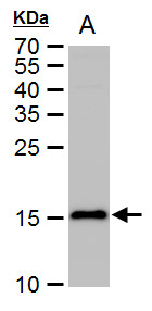
LC3B antibody detects LC3B protein by western blot analysis. A. 50 μg rat brain extract 15 % SDS-PAGE LC3B antibody dilution: 1:500Application
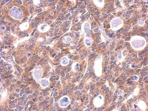
LC3B antibody detects LC3B protein at cytosol on human gastric carcinoma by immunohistochemical analysis. Sample: Paraffin-embedded gastric carcinoma. LC3B antibody dilution: 1:200.| Product Name | LC3B antibody |
|---|---|
| Antibody Type | Primary Antibodies |
| Product description | Rabbit Polyclonal antibody to LC3B (microtubule-associated protein 1 light chain 3 beta)may mediate MAPlA and MAPlB microtubule binding activity during development of the nervous system [RGD] |
| Immunogen | Full length rat LC3B Recombinant protein. |
| Modification | Acetylated |
| Clonality | Polyclonal |
|---|---|
| Isotype | IgG |
| Host Species | Rabbit |
| Tested Applications | IHC-PWB |
| WB:1:500~1:2000 IHC-P:1:50~1:200 |
|
| Species Reactivity | HumanMouseRat |
| Predicted Reactivity | Zebrafish, Japanese medaka, Xenopus laevis, Pig, Chicken, Rhesus Monkey, Chimpanzee, BovineZebrafish (91%), Japanese medaka (88%), Xenopus laevis (93%), Pig (95%), Chicken (92%), Rhesus Monkey (96%), Chimpanzee (96%), Bovine (95%) |
| Concentration | 1mg/ml (Please refer to the vial label for the specific concentration) |
| Purification | Affinity purified |
| Gene Synonyms | microtubule-associated proteins 1A/1B light chain 3 antibody autophagy-related ubiquitin-like modifier LC3 B antibody MAP1A/MAP1B LC3 B antibody MAP1A/MAP1B light chain 3 B antibody MAP1A/1B light chain 3 B antibody MAP1 light chain 3-like protein 2 a |
|---|---|
| Molecular Weight(MW) | 16 kDa (note) |
| Cellular Localization | Cytoplasm , Endomembrane system; Lipid-anchor , Cytoplasmic vesicle , autophagosome membrane; Lipid-anchor |
Application
Sample (50 ug of whole cell lysate) A: Huh7 B: virus infected Huh7 12% SDS PAGE diluted at 1:1000
Application
Western blot analysis of LC3 protein in PC-3 cell line. (1) 20ug of whole cell lysate was separated by using Urea gel (6M Urea in 13.5% SDS gel) and detected with unpurified serum diluted at 1:4000. (2) PC-3 cells were cultured with serum-containing medium (A) and serum-free medium (B) for 3 days. 5ug of whole cell lysate was separated by using Urea gel (6M Urea in 13.5% SDS gel) and detected with antibody diluted at 1:5000.
Application
LC3B antibody detects LC3B protein by western blot analysis. Various whole cell extracts (30 μg) were separated by 15 % SDS-PAGE, and blotted with LC3B antibody diluted by 1:500
Application
LC3B antibody detects LC3B protein by western blot analysis. A. 30 μg U87-MG whole cell lysate/extract B. 30 μg SK-N-SH whole cell lysate/extract 15 % SDS-PAGE LC3B antibody dilution: 1:500
Application
LC3B antibody detects LC3B protein by western blot analysis. A. 50 μg mouse brain lysate/extract 15 % SDS-PAGE LC3B antibody dilution: 1:500
Application
LC3B antibody detects LC3B protein by western blot analysis. A. 50 μg rat brain extract 15 % SDS-PAGE LC3B antibody dilution: 1:500
Application
LC3B antibody detects LC3B protein at cytosol on human gastric carcinoma by immunohistochemical analysis. Sample: Paraffin-embedded gastric carcinoma. LC3B antibody dilution: 1:200.| Positive Control | U87-MG , SK-N-SH , IMR32 , SK-N-AS , PC-3 , U87-MG , SK-N-SH , Rat brain , mouse brain |
|---|---|
| Application Notes | WB:1:500~1:2000 IHC-P:1:50~1:200 |
| Form | Liquid |
|---|---|
| Storage Instructions | Keep as concentrated solution. Aliquot and store at -20ºC or below. Avoid multiple freeze-thaw cycles. |
| Storage Buffer | 1XPBS, 20% Glycerol (pH7). 0.01% Thimerosal was added as a preservative. |


