Application
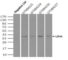
Immunoprecipitation (IP) of LDHA by using TrueMab monoclonal anti-LDHA antibodies (Negative control: IP without adding anti-LDHA antibody.). For each experiment, 500ul of DDK tagged LDHA overexpression lysates (at 1:5 dilution with HEK293T lysate), 2ug of anti-LDHA antibody and 20ul (0.1mg) of goat anti-mouse conjugated magnetic beads were mixed and incubated overnight. After extensive wash to remove any non-specific binding, the immuno-precipitated products were analyzed with rabbit anti-DDK polyclonal antibody.Application
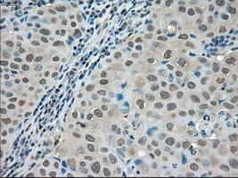
Immunohistochemical staining of paraffin-embedded Adenocarcinoma of breast tissue using anti-LDHA mouse monoclonal antibody. ( Dilution 1:50)Application
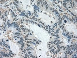
Immunohistochemical staining of paraffin-embedded Adenocarcinoma of colon tissue using anti-LDHAmouse monoclonal antibody. ( Dilution 1:50)Application
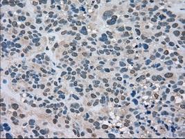
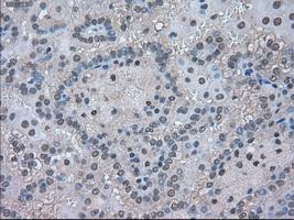
Immunohistochemical staining of paraffin-embedded Carcinoma of bladder tissue using anti-LDHAmouse monoclonal antibody. ( Dilution 1:50)Application
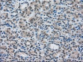
Immunohistochemical staining of paraffin-embedded Carcinoma of kidney tissue using anti-LDHAmouse monoclonal antibody. ( Dilution 1:50)Application
Immunohistochemical staining of paraffin-embedded Carcinoma of thyroid tissue using anti-LDHAmouse monoclonal antibody. ( Dilution 1:50)Application
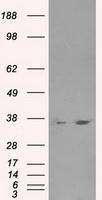
HEK293T cells were transfected with the pCMV6-ENTRY control (Left lane) or pCMV6-ENTRY LDHA (Right lane) cDNA for 48 hrs and lysed. Equivalent amounts of cell lysates (5 ug per lane) were separated by SDS-PAGE and immunoblotted with anti-LDHA.| Product Name | LDHA antibody |
|---|---|
| Antibody Type | Primary Antibodies |
| Product description | Mouse monoclonal antibody [5G10] to LDHAThe protein encoded by this gene catalyzes the conversion of L-lactate and NAD to pyruvate and NADH in the final step of anaerobic glycolysis. The protein is found predominantly in muscle tissue and belongs to the lactate dehydrogenase family. Mutations in this gene have been linked to exertional myoglobinuria. Multiple transcript variants encoding different isoforms have been found for this gene. The human genome contains several non-transcribed pseudogenes of this gene. [provided by RefSeq] |
| Immunogen | Protein expressed in 293T cell transfected with human LDHA expression vector |
| Clonality | Monoclonal |
|---|---|
| Isotype | IgG1 |
| Clone Number | 5G10 |
| Host Species | Mouse |
| Tested Applications | IHCIHC-PIPWB |
| IHC 1:50 : |
|
| Species Reactivity | Human |
| Concentration | 1mg/ml |
| Purification | Affinity purified |
| Gene Synonyms | LDHA LDH1 GSD11 LDHM |
|---|---|
| Target | LDHA |
| Tissue Specificity | is specific for human LDHA. |
Application
Immunoprecipitation (IP) of LDHA by using TrueMab monoclonal anti-LDHA antibodies (Negative control: IP without adding anti-LDHA antibody.). For each experiment, 500ul of DDK tagged LDHA overexpression lysates (at 1:5 dilution with HEK293T lysate), 2ug of anti-LDHA antibody and 20ul (0.1mg) of goat anti-mouse conjugated magnetic beads were mixed and incubated overnight. After extensive wash to remove any non-specific binding, the immuno-precipitated products were analyzed with rabbit anti-DDK polyclonal antibody.
Application
Immunohistochemical staining of paraffin-embedded Adenocarcinoma of breast tissue using anti-LDHA mouse monoclonal antibody. ( Dilution 1:50)
Application
Immunohistochemical staining of paraffin-embedded Adenocarcinoma of colon tissue using anti-LDHAmouse monoclonal antibody. ( Dilution 1:50)
Application
Immunohistochemical staining of paraffin-embedded Carcinoma of bladder tissue using anti-LDHAmouse monoclonal antibody. ( Dilution 1:50)
Application
Immunohistochemical staining of paraffin-embedded Carcinoma of kidney tissue using anti-LDHAmouse monoclonal antibody. ( Dilution 1:50)
Application
Immunohistochemical staining of paraffin-embedded Carcinoma of thyroid tissue using anti-LDHAmouse monoclonal antibody. ( Dilution 1:50)
Application
HEK293T cells were transfected with the pCMV6-ENTRY control (Left lane) or pCMV6-ENTRY LDHA (Right lane) cDNA for 48 hrs and lysed. Equivalent amounts of cell lysates (5 ug per lane) were separated by SDS-PAGE and immunoblotted with anti-LDHA.| Application Notes | IHC 1:50 : |
|---|
| Form | Liquid |
|---|---|
| Storage Instructions | For short-term storage, store at 4°C or aliquot into working amounts and store at -20°C. For long-term storage, store at -70°C (aliquotted). Avoid repeated freeze-thaw cycles. |
| Storage Buffer | PBS (PH 7.3) containing 1% BSA, 50% glycerol and 0.02% sodium azide. |


