Application
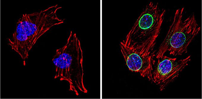
Immunofluorescent analysis of LXR beta (green) in C2C12 cells (right) compared with a negative control in the absence of primary antibody (left). Formalin-fixed cells were permeabilized with 0.1% Triton X-100 in TBS for 5-10 minutes, blocked with 3% BSA-PBS for 30 minutes at room temperature and probed with LXR beta antibody in 3% BSA-PBS at a dilution of 1:200 and incubated overnight at 4°C in a humidified chamber. Cells were washed with PBST and incubated with a proper secondary antibody. F-actin (red) was stained with a flourescent red phalloidin and nuclei (blue) were stained with DAPI. Images were taken at a magnification of 60x.Application
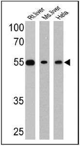
Western blot analysis of LXR beta in 25 ug of rat liver, mouse liver and HeLa lysates. Proteins were transferred to a PVDF membrane and blocked at 4°C overnight. The membrane was probed with LXR beta antibody at a dilution of 1:1000 overnight at 4°C, washed in TBST, and probed with an HRP-conjugated secondary antibody. Chemiluminescent detection was performed.| Product Name | LXR beta antibody |
|---|---|
| Antibody Type | Primary Antibodies |
| Product description | Rabbit polyclonal antibody to LXR betanuclear orphan receptor; may be involved in modulating 9-cis-retinoic acid signaling by interacting with retinoic acid receptor (RXR) [RGD, Feb 2006] |
| Immunogen | Synthetic peptide corresponding to residues R(113) Y A C R G S G T C Q M D A(126) of rat LXR beta. |
| Clonality | Polyclonal |
|---|---|
| Isotype | IgG |
| Host Species | Rabbit |
| Tested Applications | ICC/IFWB |
| ICC/IF: 1:200 - 1:1000 | |
| Species Reactivity | HumanMouseRat |
| Concentration | 1mg/ml (Please refer to the vial label for the specific concentration) |
| Purification | Affinity purified |
| Gene Synonyms | nuclear receptor subfamily 1, group H, member 2 , OR-1 , Nr1h2 |
|---|---|
| Target | LXR beta |
Application
Immunofluorescent analysis of LXR beta (green) in C2C12 cells (right) compared with a negative control in the absence of primary antibody (left). Formalin-fixed cells were permeabilized with 0.1% Triton X-100 in TBS for 5-10 minutes, blocked with 3% BSA-PBS for 30 minutes at room temperature and probed with LXR beta antibody in 3% BSA-PBS at a dilution of 1:200 and incubated overnight at 4°C in a humidified chamber. Cells were washed with PBST and incubated with a proper secondary antibody. F-actin (red) was stained with a flourescent red phalloidin and nuclei (blue) were stained with DAPI. Images were taken at a magnification of 60x.
Application
Western blot analysis of LXR beta in 25 ug of rat liver, mouse liver and HeLa lysates. Proteins were transferred to a PVDF membrane and blocked at 4°C overnight. The membrane was probed with LXR beta antibody at a dilution of 1:1000 overnight at 4°C, washed in TBST, and probed with an HRP-conjugated secondary antibody. Chemiluminescent detection was performed.| Application Notes | ICC/IF: 1:200 - 1:1000 |
|---|
| Form | Liquid |
|---|---|
| Storage Instructions | -20° C, Avoid Freeze/Thaw Cycles |
| Storage Buffer | PBS containing 0.05 % sodium azide and 1 mg/ml BSA |


