Application
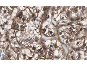
GeneTex Affinity Purified anti-MAD2L2 antibody shows strong nuclear and cytoplasmic staining of tumor cells in cancerous human kidney tissue. Tissue was formalin-fixed and paraffin embedded. Brown color indicates presence of protein, blue color shows cell nuclei.Application
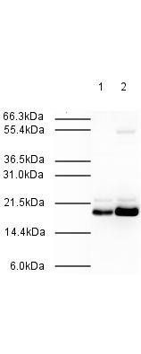
Affinity Purified Rabbit anti-MAD2L2 was used at a 1:500 dilution to detect human MAD2L2 by western blot. Both HeLa whole cell lysate (lane 1) and nuclear lysate (lane 2) were probed using this antibody. This antibody clearly detects a ~20 kDa band corresponding to human MAD2L2 (predicted molecular weight is 24 kDa). Approximately 20 µg of each lysate was loaded on a 10% SDS-PAGE. Primary antibody was reacted with the membrane at room temperature for 1 h. After subsequent washing, a 1:2,000 dilution of HRP conjugated Gt-a-Rabbit IgG was used for visualization. Exposure time was 30 sec.| Product Name | MAD2L2 antibody |
|---|---|
| Antibody Type | Primary Antibodies |
| Product description | Rabbit Polyclonal antibody to MAD2L2MAD2L2 is a component of the mitotic spindle assembly checkpoint that prevents the onset of anaphase until all chromosomes are properly aligned at the metaphase plate. MAD2L2 is a homolog of MAD2L1. |
| Immunogen | Synthetic peptide: TLTRQDLNFGQV, corresponding to amino acids 3-14 of Human Mad2L2. |
| Clonality | Polyclonal |
|---|---|
| Isotype | IgG |
| Host Species | Rabbit |
| Tested Applications | ELISAIHC-PWB |
| *Optimal dilutions/concentrations should be determined by the researcher. | |
| Species Reactivity | Human |
| Concentration | 1mg/ml |
| Purification | Affinity purified |
| Gene Synonyms | MAD2B , REV7 , POLZ2 |
|---|---|
| Target | MAD2L2 |
| Cellular Localization | Nuclear |
Application
GeneTex Affinity Purified anti-MAD2L2 antibody shows strong nuclear and cytoplasmic staining of tumor cells in cancerous human kidney tissue. Tissue was formalin-fixed and paraffin embedded. Brown color indicates presence of protein, blue color shows cell nuclei.
Application
Affinity Purified Rabbit anti-MAD2L2 was used at a 1:500 dilution to detect human MAD2L2 by western blot. Both HeLa whole cell lysate (lane 1) and nuclear lysate (lane 2) were probed using this antibody. This antibody clearly detects a ~20 kDa band corresponding to human MAD2L2 (predicted molecular weight is 24 kDa). Approximately 20 µg of each lysate was loaded on a 10% SDS-PAGE. Primary antibody was reacted with the membrane at room temperature for 1 h. After subsequent washing, a 1:2,000 dilution of HRP conjugated Gt-a-Rabbit IgG was used for visualization. Exposure time was 30 sec.| Application Notes | *Optimal dilutions/concentrations should be determined by the researcher. |
|---|
| Form | Liquid |
|---|---|
| Storage Instructions | Store vial at -20° C prior to opening. Aliquot contents and freeze at -20° C or below for extended storage. Avoid cycles of freezing and thawing. |
| Storage Buffer | 0.02 M Potassium Phosphate, 0.15 M Sodium Chloride, pH 7.2 and 0.01% (w/v) Sodium Azide |


