Application
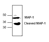
Western blot analysis of MAP-1 expression in mouse small intestine tissue lysate| Product Name | MAP-1 antibody |
|---|---|
| Antibody Type | Primary Antibodies |
| Product description | Rabbit polyclonal antibody to MAP-1Human MAP-1 (Modulator of Apoptosis Protein) is a novel Bax-associating protein. MAP-1 mediates caspase-dependent apoptosis in mammalian cells when overexpressed. MAP-1 homodimerizes and associates with the proapoptotic Bax and the prosurvival Bcl-2 and Bcl-XL of the Bcl-2 family. |
| Clonality | Polyclonal |
|---|---|
| Isotype | IgG |
| Host Species | Rabbit |
| Tested Applications | WB |
| Western blotting (1-4 µg/ml). However, the optimal concentrations should be determined individually. Jurkat cell lysate and mouse small intestine tissue lysate can be used as positive controls. The antibody recognizes 39 kDa MAP-1 from samples of human, mouse and rat origins. Reactivity to other species has not been determined. In human samples, an additional ~78 kDa dimer can be detected in Jurkat cell lysate. In mouse sample, an additional ~33 kDa cleavage fragment can also be detected in mouse small intestine tissue lysate. | |
| Species Reactivity | HumanMouse |
| Concentration | 0.5mg/ml |
| Purification | Affinity purified |
| Gene Synonyms | MAP1A, FLJ77111, MTAP1A, MAP1L, MAP1 |
|---|---|
| Target | MAP-1 |
| Tissue Specificity | This antibody is specific for MAP-1. |
Application
Western blot analysis of MAP-1 expression in mouse small intestine tissue lysate| Application Notes | Western blotting (1-4 µg/ml). However, the optimal concentrations should be determined individually. Jurkat cell lysate and mouse small intestine tissue lysate can be used as positive controls. The antibody recognizes 39 kDa MAP-1 from samples of human, mouse and rat origins. Reactivity to other species has not been determined. In human samples, an additional ~78 kDa dimer can be detected in Jurkat cell lysate. In mouse sample, an additional ~33 kDa cleavage fragment can also be detected in mouse small intestine tissue lysate. |
|---|
| Form | Liquid |
|---|---|
| Storage Instructions | The antibody solution should be gently mixed before use. Stored at -20 °C for future use. |
| Storage Buffer | PBS (pH 7.2) containing 30% glycerol, 0.5% BSA, and 0.01% thimerosal |


