Application
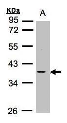
Sample (30μg whole cell lysate) A:Raji 10% SDS PAGE diluted at 1:1000Application
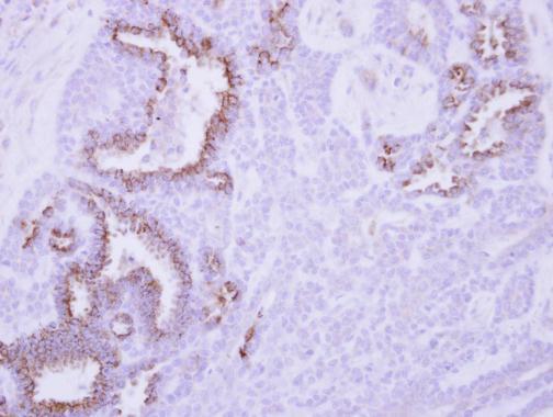
Immunohistochemical analysis of paraffin-embedded human breast cancer, using MCL1 antibody at 1:250 dilution.Application
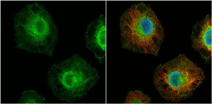
MCL1 antibody detects MCL1 protein at cytoplasm and nucleus by immunofluorescent analysis. Sample: HeLa cells were fixed in 4% paraformaldehyde at RT for 15 min. Green: MCL1 protein stained by MCL1 antibody diluted at 1:200. Red: alpha Tubulin, a cytoskeleton marker, stained by alpha Tubulin antibody [B-5-1-2] diluted at 1:10000. Blue: Hoechst 33342 staining.Application
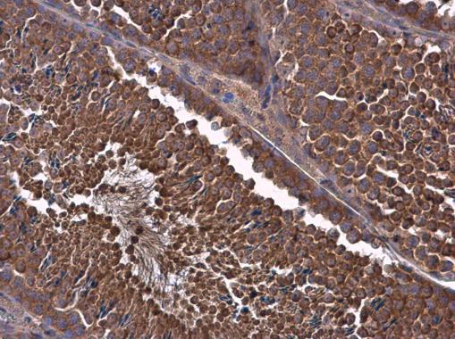
MCL1 antibody detects MCL1 protein at cytoplasm in mouse testis by immunohistochemical analysis. Sample: Paraffin-embedded mouse testis. MCL1 antibody diluted at 1:500.| Product Name | MCL1 antibody |
|---|---|
| Antibody Type | Primary Antibodies |
| Product description | Rabbit Polyclonal antibody to MCL1 (myeloid cell leukemia sequence 1 (BCL2-related))The protein encoded by this gene belongs to the Bcl-2 family. Alternative splicing occurs at this locus and two transcript variants encoding distinct isoforms have been identified. The longer gene product (isoform 1) enhances cell survival by inhibiting apoptosis while the alternatively spliced shorter gene product (isoform 2) promotes apoptosis and is death-inducing. [provided by RefSeq] |
| Immunogen | Recombinant protein encompassing a sequence within the center region of human MCL1. The exact sequence is proprietary. |
| Clonality | Polyclonal |
|---|---|
| Isotype | IgG |
| Host Species | Rabbit |
| Tested Applications | ICC/IFIHC-PWB |
| *Optimal dilutions/concentrations should be determined by the researcher.
: |
|
| Species Reactivity | HumanMouse |
| Predicted Reactivity | Rat, Dog, Cat, Rhesus Monkey, BovineRat (88%), Dog (91%), Cat (91%), Rhesus Monkey (99%), Bovine (90%) |
| Concentration | 1mg/ml |
| Purification | Affinity purified |
| Gene Synonyms | BCL2L3 antibody EAT antibody MCL1-ES antibody MCL1L antibody MCL1S antibody MGC104264 antibody MGC1839 antibody Mcl-1 antibody TM antibody bcl2-L-3 antibody mcl1/EAT antibody MCL1 antibody myeloid cell leukemia ES antibody bcl-2-like protein 3 |
|---|---|
| Molecular Weight(MW) | 37 kDa (note) |
| Cellular Localization | Membrane; Single-pass membrane protein (Potential) , Cytoplasm , Mitochondrion , Nucleus , nucleoplasm |
Application
Sample (30μg whole cell lysate) A:Raji 10% SDS PAGE diluted at 1:1000
Application
Immunohistochemical analysis of paraffin-embedded human breast cancer, using MCL1 antibody at 1:250 dilution.
Application
MCL1 antibody detects MCL1 protein at cytoplasm and nucleus by immunofluorescent analysis. Sample: HeLa cells were fixed in 4% paraformaldehyde at RT for 15 min. Green: MCL1 protein stained by MCL1 antibody diluted at 1:200. Red: alpha Tubulin, a cytoskeleton marker, stained by alpha Tubulin antibody [B-5-1-2] diluted at 1:10000. Blue: Hoechst 33342 staining.
Application
MCL1 antibody detects MCL1 protein at cytoplasm in mouse testis by immunohistochemical analysis. Sample: Paraffin-embedded mouse testis. MCL1 antibody diluted at 1:500.| Positive Control | Raji |
|---|---|
| Application Notes | *Optimal dilutions/concentrations should be determined by the researcher.
: |
| Form | Liquid |
|---|---|
| Storage Instructions | Keep as concentrated solution. Aliquot and store at -20ºC or below. Avoid multiple freeze-thaw cycles. |
| Storage Buffer | 0.1M Tris, 0.1M Glycine, 10% Glycerol (pH7). 0.01% Thimerosal was added as a preservative. |


