Application
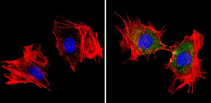
ICC/IF analysis of 3T3-L1 cells with (right) or without (left) using MDC1 antibody at a dilution of 1:100 (green). Actin was stained using Alexa Fluor 554 (red) and nuclei were stained with DAPI (blue). Images were taken at a magnification of 60x.Application
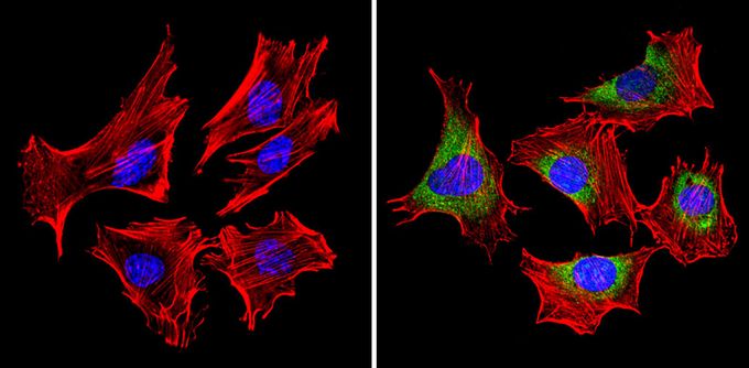
ICC/IF analysis of C2C12 cells with (right) or without (left) using MDC1 antibody at a dilution of 1:100 (green). Actin was stained using Alexa Fluor 554 (red) and nuclei were stained with DAPI (blue). Images were taken at a magnification of 60x.Application
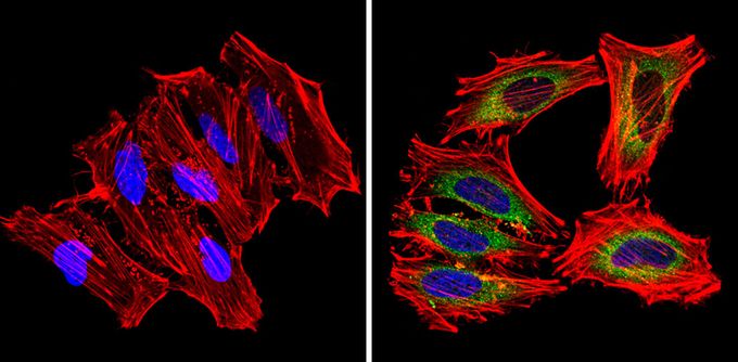
ICC/IF analysis of HeLa cells with (right) or without (left) MDC1 antibody at a dilution of 1:100 (green). Actin was stained using Alexa Fluor 554 (red) and nuclei were stained with DAPI (blue). Images were taken at a magnification of 60x.| Product Name | MDC1 antibody |
|---|---|
| Antibody Type | Primary Antibodies |
| Product description | Rabbit Polyclonal antibody to MDC1The mitochondrial dicarboxylate carrier protein (mDIC) belongs to superfamily of mitochondrial transporters. It has been demonstrated that the expression of mDIC leads to hyperpolarization of the mitochondria, as well as succinate uptake by the mitochondria. It is highly expressed in adipose tissue, where its expression is downregulated by insulin and upregulated by free fatty acids. mDIC is also thought to play a role in glyceroneogenesis. |
| Immunogen | Synthetic peptide corresponding to the C-terminal residues L(276) R K H F G I K V P T T(287) of mDIC. |
| Clonality | Polyclonal |
|---|---|
| Host Species | Rabbit |
| Tested Applications | ICC/IFWB |
| ICC/IF: 1:50 - 1:500: |
|
| Species Reactivity | HumanMouse |
| Concentration | 1mg/ml |
| Purification | Affinity purified |
| Gene Synonyms | Slc25a10 Dic solute carrier family 25 (mitochondrial carrier dicarboxylate transporter) member 10 |
|---|---|
| Target | MDC1 |
Application
ICC/IF analysis of 3T3-L1 cells with (right) or without (left) using MDC1 antibody at a dilution of 1:100 (green). Actin was stained using Alexa Fluor 554 (red) and nuclei were stained with DAPI (blue). Images were taken at a magnification of 60x.
Application
ICC/IF analysis of C2C12 cells with (right) or without (left) using MDC1 antibody at a dilution of 1:100 (green). Actin was stained using Alexa Fluor 554 (red) and nuclei were stained with DAPI (blue). Images were taken at a magnification of 60x.
Application
ICC/IF analysis of HeLa cells with (right) or without (left) MDC1 antibody at a dilution of 1:100 (green). Actin was stained using Alexa Fluor 554 (red) and nuclei were stained with DAPI (blue). Images were taken at a magnification of 60x.| Application Notes | ICC/IF: 1:50 - 1:500: |
|---|
| Form | Liquid |
|---|---|
| Storage Instructions | Keep as concentrated solution. Aliquot and store at -20ºC or below. Avoid multiple freeze-thaw cycles. |
| Storage Buffer | Antisera containing 0.05% sodium azide. |


