Application
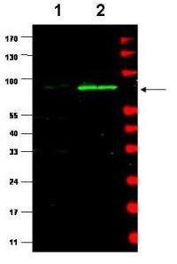
Western blot using GeneTex's affinity purified Anti-MDM2 (Rabbit) is shown to detect a band (arrow) corresponding to mouse MDM2 protein present in mouse MEF cells (lane 2) but not human kidney HEK293 cells (lane 1). Approximately 35 ug of lysate was separated by 4-20% Tris Glycine SDS-PAGE. After blocking the membrane with 5% normal goat serum, 0.5% BLOTTO in PBS, the membrane was probed for overnight at 4° with the primary antibody diluted to 1:500 in 1% normal goat serum, 0.1% BLOTTO in PBS. The membrane was washed and reacted with a 1:10,000 dilution of IRDye800 conjugated Gt-a-Rabbit IgG [H&L] for 45 min at room temperature (800 nm channel, green). Molecular weight estimation was made by comparison to prestained MW markers indicated at the right (700 nm channel, red). IRDye800 fluorescence image was captured using the Odyssey® Infrared Imaging System developed by LI-COR. IRDye is a trademark of LI-COR, Inc. Other detection systems will yield similar results.| Product Name | MDM2 antibody |
|---|---|
| Antibody Type | Primary Antibodies |
| Product description | Rabbit Polyclonal antibody to MDM2MDM2 is a nuclear phosphoprotein with an apparent molecular mass of 90 kD that forms a complex with the p53 tumor suppressor protein. Human MDM2 was identified as a homologous product of the 'murine double minute 2' gene (mdm2). The MDM2 gene enhances the tumorigenic potential of cells when it is overexpressed and encodes a putative transcription factor. Forming a tight complex with the p53 gene, the MDM2 oncogene can inhibit p53-mediated transactivation. MDM2 binds to p53 and amplification of MDM2 in sarcomas leads to escape from p53-regulated growth control. This mechanism of tumorigenesis parallels that for virus-induced tumors in which viral oncogene products bind to and functionally inactivate p53.Over-expression of the |
| Immunogen | This affinity purified antibody was prepared from whole rabbit serum produced by repeated immunizations with a synthetic peptide corresponding to aa 177-195 of mouse MDM2. |
| Clonality | Polyclonal |
|---|---|
| Isotype | IgG |
| Host Species | Rabbit |
| Tested Applications | ELISAIPWB |
| *Optimal dilutions/concentrations should be determined by the researcher.
: |
|
| Species Reactivity | Mouse |
| Concentration | 1mg/ml |
| Purification | Affinity purified |
| Gene Synonyms | 1700007J15RIK AA415488 |
|---|---|
| Target | MDM2 |
| Cellular Localization | Colocalizes with RASSF1 isoform A in the nucleus (By similarity). Expressed predominantly in the nucleoplasm. Interaction with ARF(P14) results in the localization of both proteins to the nucleolus. The nucleolar localization signals in both ARF(P14) and |
Application
Western blot using GeneTex's affinity purified Anti-MDM2 (Rabbit) is shown to detect a band (arrow) corresponding to mouse MDM2 protein present in mouse MEF cells (lane 2) but not human kidney HEK293 cells (lane 1). Approximately 35 ug of lysate was separated by 4-20% Tris Glycine SDS-PAGE. After blocking the membrane with 5% normal goat serum, 0.5% BLOTTO in PBS, the membrane was probed for overnight at 4° with the primary antibody diluted to 1:500 in 1% normal goat serum, 0.1% BLOTTO in PBS. The membrane was washed and reacted with a 1:10,000 dilution of IRDye800 conjugated Gt-a-Rabbit IgG [H&L] for 45 min at room temperature (800 nm channel, green). Molecular weight estimation was made by comparison to prestained MW markers indicated at the right (700 nm channel, red). IRDye800 fluorescence image was captured using the Odyssey® Infrared Imaging System developed by LI-COR. IRDye is a trademark of LI-COR, Inc. Other detection systems will yield similar results.| Application Notes | *Optimal dilutions/concentrations should be determined by the researcher.
: |
|---|
| Form | Liquid |
|---|---|
| Storage Instructions | Store vial at -20° C prior to opening. Aliquot contents and freeze at -20° C or below for extended storage. Avoid cycles of freezing and thawing. |
| Storage Buffer | 0.02 M Potassium Phosphate, 0.15 M Sodium Chloride, pH 7.2 and 0.01% (w/v) Sodium Azide |


