Application
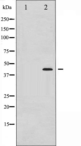
Western blot analysis of MEK1/2 phosphorylation expression in UV treated Jurkat whole cell lysates,The lane on the left is treated with the antigen-specific peptide.WB
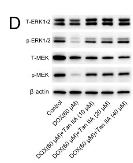
(D) Representative images showing that the protein levels of p-MEK and p-ERK1/2 were increased by Tan IIA as determined by Western blot.WB
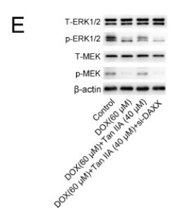
(E) The protein expression of p-ERK1/2 and p-MEK was measured by Western blotting.WB
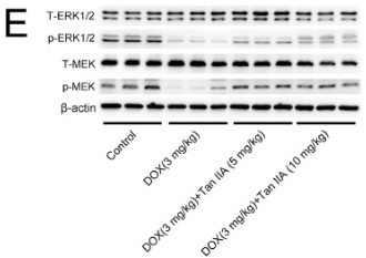
(E) Representative images showing the protein levels of p-MEK and p-ERK1/2.IHC
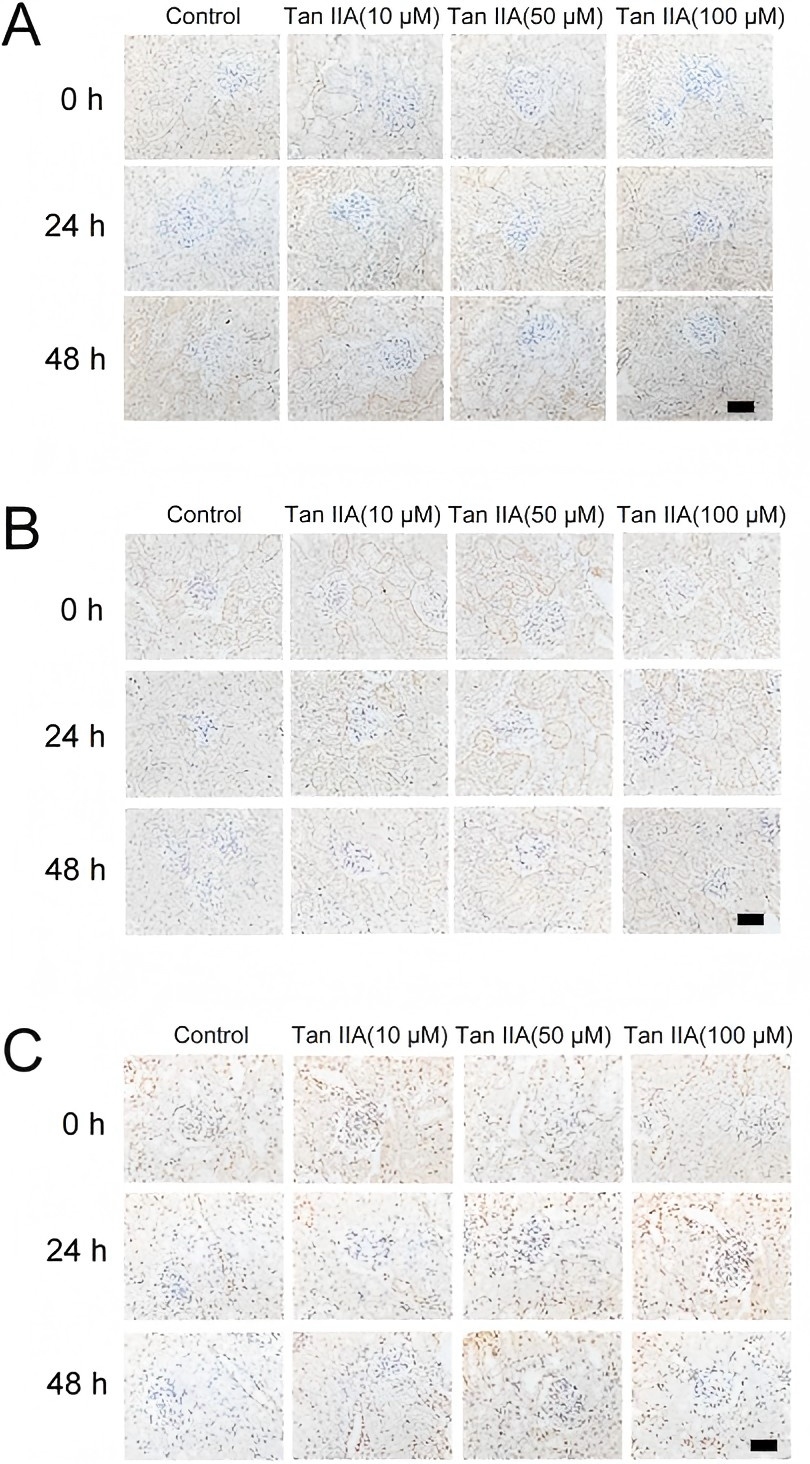
Tan IIA increased the expression of p-MEK (A), p-ERK1/2 (B) and p-GSK-3β (C), as assessed by immunohistochemical analysis.WB
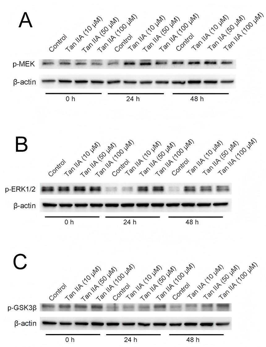
Tan IIA increases the expression of p-MEK (A), p-ERK1/2 (B) and p-GSK-3β (C), as detected by immunoblotting.WB
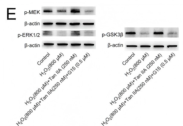
(E) Tan IIA increased the expression of p-MEK, p-ERK1/2 and p-GSK-3β, as detected by immunoblotting. G15 could diminish the expression of these proteins induced by Tan IIA.| Product Name | MEK1/2 (phospho Ser221) antibody |
|---|---|
| Antibody Type | Primary Antibodies |
| Product description | Rabbit Polyclonal antibody to MEK1/2 (phospho Ser221)The protein encoded by this gene is a member of the dual specificity protein kinase family, which acts as a mitogen-activated protein (MAP) kinase kinase. MAP kinases, also known as extracellular signal-regulated kinases (ERKs), act as an integration point for multiple biochemical signals. This protein kinase lies upstream of MAP kinases and stimulates the enzymatic activity of MAP kinases upon wide variety of extra- and intracellular signals. As an essential component of MAP kinase signal transduction pathway, this kinase is involved in many cellular processes such as proliferation, differentiation, transcription regulation and development. [provided by RefSeq, Jul 2008] |
| Immunogen | A synthesized peptide derived from human MEK1/2 around the phosphorylation site of Ser221 |
| Modification | p-Ser221 |
| Clonality | Polyclonal |
|---|---|
| Isotype | IgG |
| Host Species | Rabbit |
| Tested Applications | IHCWB |
| WB 1:500-1:2000, IHC 1:50-1:200: |
|
| Species Reactivity | HumanMouseRat |
| Concentration | 1mg/ |
| Purification | Affinity purified |
| Gene Synonyms | MEK1/2 (phospho Ser221) S221 Phospho MEK1/2 (phospho Ser221) Phospho S221 MEK1/2 (phospho Ser221) (pSer221) phospho MEK1/2 (phospho Ser221) (pS221) MEK1/2 (phospho Ser221) (phospho Ser221) phospho MEK1/2 ( |
|---|---|
| Target | MEK1/2 (phospho Ser221) |
| Tissue Specificity | MAP2K1 |
Application
Western blot analysis of MEK1/2 phosphorylation expression in UV treated Jurkat whole cell lysates,The lane on the left is treated with the antigen-specific peptide.
WB
(D) Representative images showing that the protein levels of p-MEK and p-ERK1/2 were increased by Tan IIA as determined by Western blot.
WB
(E) The protein expression of p-ERK1/2 and p-MEK was measured by Western blotting.
WB
(E) Representative images showing the protein levels of p-MEK and p-ERK1/2.
IHC
Tan IIA increased the expression of p-MEK (A), p-ERK1/2 (B) and p-GSK-3β (C), as assessed by immunohistochemical analysis.
WB
Tan IIA increases the expression of p-MEK (A), p-ERK1/2 (B) and p-GSK-3β (C), as detected by immunoblotting.
WB
(E) Tan IIA increased the expression of p-MEK, p-ERK1/2 and p-GSK-3β, as detected by immunoblotting. G15 could diminish the expression of these proteins induced by Tan IIA.| Application Notes | WB 1:500-1:2000, IHC 1:50-1:200: |
|---|
| Form | Liquid |
|---|---|
| Storage Instructions | Keep as concentrated solution. Aliquot and store at -20ºC or below. Avoid multiple freeze-thaw cycles. |
| Storage Buffer | phosphate buffered saline , pH 7.4, 150mM NaCl, 0.02% sodium azide and 50% glycerol. |


