Application
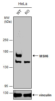
Wild-type (WT) and MSH6 knockout (KO) HeLa cell extracts (30 µg) were separated by 5% SDS-PAGE, and the membrane was blotted with MSH6 antibody [C1C2], Internal diluted at 1:1000. The HRP-conjugated anti-rabbit IgG antibody was used to detect the primary antibody.Application
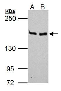
MSH6 antibody [C1C2], Internal detects MSH6 protein by Western blot analysis. A. 30 µg Neuro2A whole cell lysate/extract B. 30 µg GL261 whole cell lysate/extract 5% SDS-PAGE MSH6 antibody [C1C2], Internal dilution: 1:1000Application
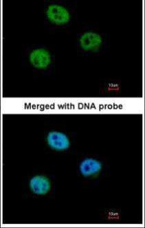
Immunofluorescence analysis of paraformaldehyde-fixed HeLa, using GTBP (MSH6) antibody at 1:500 dilution.Application
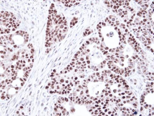
Immunohistochemical analysis of paraffin-embedded NCIN87 Xenograft, using GTBP (MSH6) antibody at 1:100 dilution.Application
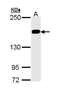
Sample (30 ug of whole cell lysate) A: Hep G2 5% SDS PAGE diluted at 1:1000Application
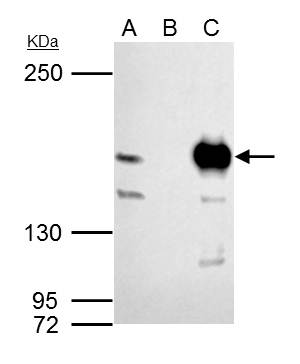
MSH6 antibody [C1C2], Internal immunoprecipitates MSH6 protein in IP experiments. IP samples: 293T whole cell extract A. 40 μg 293T whole cell extract B. Control with 4 μg of preimmune Rabbit IgG C. Immunoprecipitation of MSH6 protein by 4 μg MSH6 antibody [C1C2], Internal 5 % SDS-PAGE The immunoprecipitated MSH6 protein was detected by MSH6 antibody [C1C2], Internal diluted at 1:1000. [EasyBlot anti-rabbit IgG was used as a secondary reagent]| Product Name | MSH6 antibody [C1C2], Internal |
|---|---|
| Antibody Type | Primary Antibodies |
| Product description | Rabbit Polyclonal antibody to MSH6 (mutS homolog 6 (E. coli))This gene encodes a protein similar to the MutS protein. In E. coli, the MutS protein helps in the recognition of mismatched nucleotides, prior to their repair. A highly conserved region of approximately 150 aa, called the Walker-A adenine nucleotide binding motif, exists in MutS homologs. The encoded protein of this gene combines with MSH2 to form a mismatch recognition complex that functions as a bidirectional molecular switch that exchanges ADP and ATP as DNA mismatches are bound and dissociated. Mutations in this gene have been identified in individuals with hereditary nonpolyposis colon cancer (HNPCC) and endometrial cancer. [provided by RefSeq] |
| Immunogen | Recombinant protein encompassing a sequence within the center region of human MSH6. The exact sequence is proprietary. |
| Clonality | Polyclonal |
|---|---|
| Isotype | IgG |
| Host Species | Rabbit |
| Tested Applications | EMSAICC/IFIHC-PIPWB |
| *Optimal dilutions/concentrations should be determined by the researcher. | |
| Species Reactivity | HumanMouseZebra Fish |
| Predicted Reactivity | Rhesus Monkey, BovineRhesus Monkey (99%), Bovine (93%) |
| Concentration | 1mg/ml (Please refer to the vial label for the specific concentration) |
| Purification | Affinity purified |
| Gene Synonyms | GTBP antibody, HNPCC5 antibody, HSAP antibody, MSH6 antibody, DNA mismatch repair protein Msh6 antibody, mutS-alpha 160 kDa subunit antibody, GTMBP antibody, sperm-associated protein antibody, p160 antibody, hMSH6 antibody, G/T mismatch-binding protein ant |
|---|---|
| Molecular Weight(MW) | 153 kDa (note) (4) |
| Cellular Localization | Nucleus |
Application
Wild-type (WT) and MSH6 knockout (KO) HeLa cell extracts (30 µg) were separated by 5% SDS-PAGE, and the membrane was blotted with MSH6 antibody [C1C2], Internal diluted at 1:1000. The HRP-conjugated anti-rabbit IgG antibody was used to detect the primary antibody.
Application
MSH6 antibody [C1C2], Internal detects MSH6 protein by Western blot analysis. A. 30 µg Neuro2A whole cell lysate/extract B. 30 µg GL261 whole cell lysate/extract 5% SDS-PAGE MSH6 antibody [C1C2], Internal dilution: 1:1000
Application
Immunofluorescence analysis of paraformaldehyde-fixed HeLa, using GTBP (MSH6) antibody at 1:500 dilution.
Application
Immunohistochemical analysis of paraffin-embedded NCIN87 Xenograft, using GTBP (MSH6) antibody at 1:100 dilution.
Application
Sample (30 ug of whole cell lysate) A: Hep G2 5% SDS PAGE diluted at 1:1000
Application
MSH6 antibody [C1C2], Internal immunoprecipitates MSH6 protein in IP experiments. IP samples: 293T whole cell extract A. 40 μg 293T whole cell extract B. Control with 4 μg of preimmune Rabbit IgG C. Immunoprecipitation of MSH6 protein by 4 μg MSH6 antibody [C1C2], Internal 5 % SDS-PAGE The immunoprecipitated MSH6 protein was detected by MSH6 antibody [C1C2], Internal diluted at 1:1000. [EasyBlot anti-rabbit IgG was used as a secondary reagent]| Positive Control | HepG2 , Neuro2A , GL261 |
|---|---|
| Application Notes | *Optimal dilutions/concentrations should be determined by the researcher. |
| Form | Liquid |
|---|---|
| Storage Instructions | Keep as concentrated solution. Aliquot and store at -20ºC or below. Avoid multiple freeze-thaw cycles. |
| Storage Buffer | 0.1M Tris, 0.1M Glycine, 10% Glycerol (pH7). 0.01% Thimerosal was added as a preservative. |


