Application
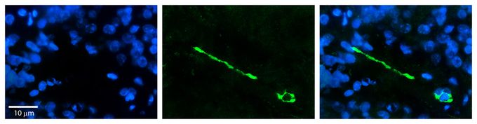
IHC-P analysis of human pineal tissue using MTUS1 antibody at 1:100. Left to right:DAPI, MTUS1, Merge. The low pH, heat-induced antigen retrieval method utilizing Sodium Citrate buffer was performed.Application
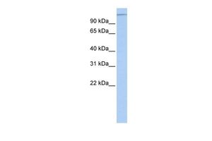
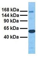
WB analysis of HeLa cells using MTUS1 antibody at 0.2-1μg/ml.Application
WB analysis of human ovary tumor tissue using MTUS1 antibody at 1μg/ml.Application
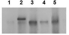
WB analysis of human MCF-7, HeLa, and human cancer cell lines using MTUS1 antibody at 1:2000. Lane : 1 : MCF-7 cells 2 : MCF-7 cells over-expressing GFP tagged MTUS1 protein 3 : HeLa cells 4 and 5 : Human cancer cell lines The observed size of endogenous MTUS1 protein is at ~180kDa, and GFP tagged MTSU1 protein is at ~210kDa.Application
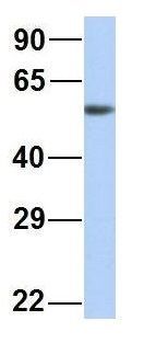
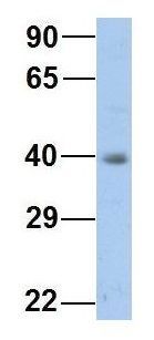
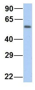
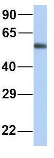
WB analysis of human placenta tissue using MTUS1 antibody at 1μg/ml.Application
WB analysis of HepG2 cells using MTUS1 antibody at 1μg/ml.Application
WB analysis of human fetal heart tissue using MTUS1 antibody at 1μg/ml.Application
WB analysis of human fetal lung tissue using MTUS1 antibody at 1μg/ml.| Product Name | MTUS1 antibody, Internal |
|---|---|
| Antibody Type | Primary Antibodies |
| Product description | Rabbit Polyclonal antibody to MTUS1This gene encodes a protein which contains a C-terminal domain able to interact with the angiotension II (AT2) receptor and a large coiled-coil region allowing dimerization. Multiple alternatively spliced transcript variants encoding different isoforms have been found for this gene. One of the transcript variants has been shown to encode a mitochondrial protein that acts as a tumor suppressor and partcipates in AT2 signaling pathways. Other variants may encode nuclear or transmembrane proteins but it has not been determined whether they also participate in AT2 signaling pathways. [provided by RefSeq, Jul 2008] |
| Immunogen | A synthetic peptide corresponding to an Internal region of Human MTUS1 |
| Clonality | Polyclonal |
|---|---|
| Isotype | IgG |
| Host Species | Rabbit |
| Tested Applications | IHC-PWB |
| *Optimal dilutions/concentrations should be determined by the researcher.
: |
|
| Species Reactivity | Human |
| Predicted Reactivity | Cow: 100%; Dog: 100%; Guinea Pig: 86%; Human: 100%; Mouse: 93%; Rabbit: 86%; Rat: 93% |
| Concentration | 1mg/ml |
| Purification | Affinity purified |
| Gene Synonyms | MTUS1 MTSG1 MP44 ATIP ICIS ATBP |
|---|---|
| Target | MTUS1 |
| Molecular Weight(MW) | 141 KDa (note) |
Application
IHC-P analysis of human pineal tissue using MTUS1 antibody at 1:100. Left to right:DAPI, MTUS1, Merge. The low pH, heat-induced antigen retrieval method utilizing Sodium Citrate buffer was performed.
Application
WB analysis of HeLa cells using MTUS1 antibody at 0.2-1μg/ml.
Application
WB analysis of human ovary tumor tissue using MTUS1 antibody at 1μg/ml.
Application
WB analysis of human MCF-7, HeLa, and human cancer cell lines using MTUS1 antibody at 1:2000. Lane : 1 : MCF-7 cells 2 : MCF-7 cells over-expressing GFP tagged MTUS1 protein 3 : HeLa cells 4 and 5 : Human cancer cell lines The observed size of endogenous MTUS1 protein is at ~180kDa, and GFP tagged MTSU1 protein is at ~210kDa.
Application
WB analysis of human placenta tissue using MTUS1 antibody at 1μg/ml.
Application
WB analysis of HepG2 cells using MTUS1 antibody at 1μg/ml.
Application
WB analysis of human fetal heart tissue using MTUS1 antibody at 1μg/ml.
Application
WB analysis of human fetal lung tissue using MTUS1 antibody at 1μg/ml.| Positive Control | HeLa , HepG2 , human fetal heart , human fetal lung , human ovary tumor , human placenta , MCF-7 |
|---|---|
| Application Notes | *Optimal dilutions/concentrations should be determined by the researcher.
: |
| Form | Liquid |
|---|---|
| Storage Instructions | Keep as concentrated solution. For short-term storage, store at 4ºC. For long-term storage, aliquot and store at -20ºC or below. Avoid multiple freeze-thaw cycles. |
| Storage Buffer | 1x PBS buffer with 0.09% (w/v) sodium azide and 2% sucrose. |


