Application
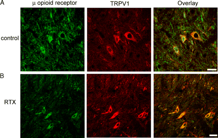
Double immunofluorescence labeling showing the presence of a subpopulation of spinal lamina II neurons that contained both µ opioid receptor (green) and TRPV1 (red) immunoreactivity in a vehicle (A)- and a RTX (B)-treated rats. Co-localization of the µ opioid receptor and TRPV1 immunoreactivity is indicated in yellow when 2 images are digitally merged. All images are single confocal optical sections. Scale bar: 20 µmApplication
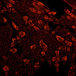
MOR staining of Rat DRG (dilution: 1:100) incubation at 4 °C overnight. Secondary antibody is anti-Rabbit Rhodamine Red (dilution:1:200) incubation at room temperature for 1 hour.| Product Name | Mu Opioid receptor antibody |
|---|---|
| Antibody Type | Primary Antibodies |
| Product description | Rabbit polyclonal to Mu Opioid ReceptorThe powerful effects of opiate are mediated through the opioid receptors located on the plasma membrane of neurons and other peripheral cells. There are at least 3 types of opioid receptors, mu, kappa, and delta, each with a distinct pharmacologic profile. The mu opioid receptor is the primary site of action for the most commonly used opioids, including morphine, heroin, fentanyl, and methadone. Three types of opioid receptors have been cloned mu, delta, and kappa. Opioid receptors are seven transmembrane G-protein coupled receptors. They share a high degree of homology and are most divergent at the N and C-termini. Activation of mu opioid receptors leads to a decrease in neuronal excitability. Expression of Mu Opioid Receptor has been reported mostly in brain and spinal cord. |
| Immunogen | Synthetic peptide: NHQLENLEAETAPLP, corresponding to C terminal amino acids 384-398 of Rat Mu Opioid Receptor. |
| Clonality | Polyclonal |
|---|---|
| Isotype | IgG |
| Host Species | Rabbit |
| Tested Applications | ICC/IFIHCIHC-FWB |
| ICC: Use at a dilution of 1/2500 - 1/5000. IHC-Fr: Use at a dilution of 1/2500 - 1/5000. WB: Use at a dilution of 1/1000. Predicted molecular weight: 48 kDa. Optimal dilutions/concentrations should be determined by the end user.: |
|
| Species Reactivity | HumanMouseMonkeyPigRatGuinea PigPrimateBovine |
| Concentration | 1mg/ml |
| Purification | Affinity purified |
| Gene Synonyms | Oprm1 MORA 25601 Mu Opioid receptor |
|---|---|
| Target | Mu Opioid receptor |
| Cellular Localization | Integral membrane protein. |
Application
Double immunofluorescence labeling showing the presence of a subpopulation of spinal lamina II neurons that contained both µ opioid receptor (green) and TRPV1 (red) immunoreactivity in a vehicle (A)- and a RTX (B)-treated rats. Co-localization of the µ opioid receptor and TRPV1 immunoreactivity is indicated in yellow when 2 images are digitally merged. All images are single confocal optical sections. Scale bar: 20 µm
Application
MOR staining of Rat DRG (dilution: 1:100) incubation at 4 °C overnight. Secondary antibody is anti-Rabbit Rhodamine Red (dilution:1:200) incubation at room temperature for 1 hour.| Application Notes | ICC: Use at a dilution of 1/2500 - 1/5000. IHC-Fr: Use at a dilution of 1/2500 - 1/5000. WB: Use at a dilution of 1/1000. Predicted molecular weight: 48 kDa. Optimal dilutions/concentrations should be determined by the end user.: |
|---|
| Form | Liquid |
|---|---|
| Storage Instructions | Aliquot and store at -20°C or -80°C. Avoid repeated freeze / thaw cycles. |
| Storage Buffer | Preservative: 0.05% Sodium Azide; Constituents: Whole Serum |


