Application
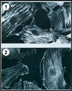
Localization of microfilament associated proteins in PTK2 cells using (1) Monoclonal Anti-Myosin (Light Chain), Clone No. MY-21 and (2) Monoclonal Anti-α-Actinin, Clone No. BM-75.2 .| Product Name | Myosin Light chain antibody |
|---|---|
| Antibody Type | Primary Antibodies |
| Product description | Mouse monoclonal [MY - 21] to Myosin light chainContractile proteins are ubiquitous in eukaryotic cells. Their presence is related to motile functions. The two major cytoskeletal proteins implicated in cell motility are actin and myosin. Numerous investigators have shown that actin and myosin are constituents of many cell types and are involved in a myriad of cellular processes including locomotion, secretion, cytoplasmic streaming, phagocytosis, and cytokinesis. Myosin is a 500,000 dalton protein known to interact with identical heavy chains (200,000 daltons each) and four light chains (15,000-20,000 daltons). Myosin molecules consist of two major regions: tail (rod) and head regions. Myosin has three major biological functions:1. Myosin molecules spontaneously assemble into filaments in solutions of physiologic ionic strength and pH. Thick filaments consist mainly of myosin molecules.2. Myosin is an enzyme (ATPase) and its activity is the immediate source of the free energy that drives muscle contraction.3. Myosin binds to the polymerized form of actin, the major constituent of the thin filament.Myosin can be viewed in immunofluorescent microscopy enabling the delineation of the intracellular organization of proteins that are in the form of a supramolecular structure. |
| Immunogen | Chicken lens membranes. |
| Clonality | Monoclonal |
|---|---|
| Isotype | IgM |
| Clone Number | MY-21 |
| Host Species | Mouse |
| Tested Applications | DotICC/IFWB |
| Dot: Use at an assay dependent dilution. The antibody reacts with purified myosin light chains (20,000 daltons) in both the native protein state or an SDS-heat denatured, reduced state. IF: Use at a dilution of 1/200. The antibody localizes an epitope on stress fibers of well spread cells. WB: Use at an assay dependent dilution. Detects a band of approximately 20 kDa. Monoclonal Anti-Myosin reacts best with the 20,000 dalton light chains of chicken gizzard, intestine or pig stomach myosin (SDS heat denatured and reduced) using an immunoblotting technique. Monoclonal Anti-Myosin shows no reactivity on frozen sections of human skeletal muscle. The product shows no reactivity with intact myosin molecules in their native state. Heat denaturation in the presence of SDS and a reducing agent (e.g. mercaptoethanol) makes accessible for the monoclonal antibody an epitope on the myosin light chains. Optimal dilutions/concentrations should be determined by the end user.: |
|
| Species Reactivity | HumanPigRabbitBovine |
| Concentration | 1mg/ml |
| Purification | Affinity purified |
| Gene Synonyms | 160780 MYL2 4632 MLC3F 416874 4633 P10916 Myosin Light chain MLC1F CMH10 MYL1 396470 160781 MLC2 P05976 |
|---|---|
| Target | Myosin Light chain |
| Cellular Localization | Cytoplasmic |
Application
Localization of microfilament associated proteins in PTK2 cells using (1) Monoclonal Anti-Myosin (Light Chain), Clone No. MY-21 and (2) Monoclonal Anti-α-Actinin, Clone No. BM-75.2 .| Application Notes | Dot: Use at an assay dependent dilution. The antibody reacts with purified myosin light chains (20,000 daltons) in both the native protein state or an SDS-heat denatured, reduced state. IF: Use at a dilution of 1/200. The antibody localizes an epitope on stress fibers of well spread cells. WB: Use at an assay dependent dilution. Detects a band of approximately 20 kDa. Monoclonal Anti-Myosin reacts best with the 20,000 dalton light chains of chicken gizzard, intestine or pig stomach myosin (SDS heat denatured and reduced) using an immunoblotting technique. Monoclonal Anti-Myosin shows no reactivity on frozen sections of human skeletal muscle. The product shows no reactivity with intact myosin molecules in their native state. Heat denaturation in the presence of SDS and a reducing agent (e.g. mercaptoethanol) makes accessible for the monoclonal antibody an epitope on the myosin light chains. Optimal dilutions/concentrations should be determined by the end user.: |
|---|
| Form | Liquid |
|---|---|
| Storage Instructions | Keep as concentrated solution. Store at 4ºC short term. For extended storage aliquot and store at -20ºC or below. Avoid freeze-thaw cycles. |
| Storage Buffer | Preservative: 15mM Sodium Azide |


