Application
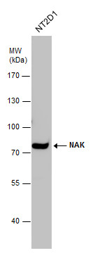
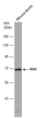
NAK antibody detects NAK protein by western blot analysis. Whole cell extracts (30 μg) was separated by 7.5% SDS-PAGE, and the membrane was blotted with NAK antibody diluted at 1:1000.Application
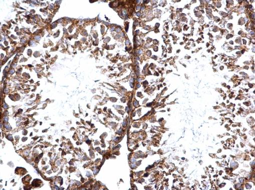
NAK antibody [N2C1], Internal detects NAK protein at cytosol and nucleus on mouse testis by immunohistochemical analysis. Sample: Paraffin-embedded mouse testis. NAK antibody [N2C1], Internal dilution: 1:500.Application
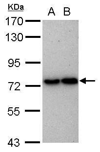
Sample (30 ug of whole cell lysate) A: HeLa B: HepG2 7.5% SDS PAGE diluted at 1:1000Application
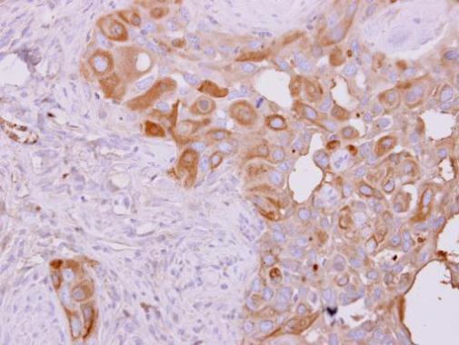
NAK antibody [N2C1], Internal detects TBK1 protein at cytoplasm on H441 xenograft by immunohistochemical analysis. Sample: Paraffin-embedded H441 xenograft. NAK antibody [N2C1], Internal dilution: 1:500.Application
Mouse tissue extract (50 µg) was separated by 7.5% SDS-PAGE, and the membrane was blotted with NAK antibody [N2C1], Internal diluted at 1:500.Application
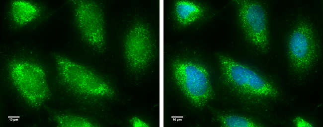
NAK antibody [N2C1], Internal detects NAK protein at cytoplasm by immunofluorescent analysis. Sample: HeLa cells were fixed in 4% paraformaldehyde for 10 min. Green: NAK protein stained by NAK antibody [N2C1], Internal diluted at 1:100. Blue: Hoechst 33342 staining. Scale bar = 10 μm.| Product Name | NAK antibody , Internal |
|---|---|
| Antibody Type | Primary Antibodies |
| Product description | Rabbit Polyclonal antibody to NAK (TANK-binding kinase 1)The NF-kappa-B (NFKB) complex of proteins is inhibited by I-kappa-B (IKB) proteins, which inactivate NFKB by trapping it in the cytoplasm. Phosphorylation of serine residues on the IKB proteins by IKB kinases marks them for destruction via the ubiquitination pathway, thereby allowing activation and nuclear translocation of the NFKB complex. The protein encoded by this gene is similar to IKB kinases and can mediate NFKB activation in response to certain growth factors. For example, the protein can form a complex with the IKB protein TANK and TRAF2 and release the NFKB inhibition caused by TANK. [provided by RefSeq] |
| Immunogen | Recombinant protein encompassing a sequence within the center region of human NAK. The exact sequence is proprietary. |
| Clonality | Polyclonal |
|---|---|
| Isotype | IgG |
| Host Species | Rabbit |
| Tested Applications | ICC/IFIHC-PWB |
| *Optimal dilutions/concentrations should be determined by the researcher.
: |
|
| Species Reactivity | HumanMouse |
| Predicted Reactivity | Rat, Pig, Chicken, Rhesus Monkey, BovineRat (93%), Pig (98%), Chicken (89%), Rhesus Monkey (98%), Bovine (96%) |
| Concentration | 1mg/ml |
| Purification | Affinity purified |
| Gene Synonyms | FLJ11330 antibody NAK antibody T2K antibody TBK1 antibody NF-kB-activating kinase antibody serine/threonine-protein kinase TBK1 antibody NF-kappa-B-activating kinase antibody TANK-binding kinase 1 antibody |
|---|---|
| Molecular Weight(MW) | 84 kDa (note) |
| Cellular Localization | Cytoplasm |
Application
NAK antibody detects NAK protein by western blot analysis. Whole cell extracts (30 μg) was separated by 7.5% SDS-PAGE, and the membrane was blotted with NAK antibody diluted at 1:1000.
Application
NAK antibody [N2C1], Internal detects NAK protein at cytosol and nucleus on mouse testis by immunohistochemical analysis. Sample: Paraffin-embedded mouse testis. NAK antibody [N2C1], Internal dilution: 1:500.
Application
Sample (30 ug of whole cell lysate) A: HeLa B: HepG2 7.5% SDS PAGE diluted at 1:1000
Application
NAK antibody [N2C1], Internal detects TBK1 protein at cytoplasm on H441 xenograft by immunohistochemical analysis. Sample: Paraffin-embedded H441 xenograft. NAK antibody [N2C1], Internal dilution: 1:500.
Application
Mouse tissue extract (50 µg) was separated by 7.5% SDS-PAGE, and the membrane was blotted with NAK antibody [N2C1], Internal diluted at 1:500.
Application
NAK antibody [N2C1], Internal detects NAK protein at cytoplasm by immunofluorescent analysis. Sample: HeLa cells were fixed in 4% paraformaldehyde for 10 min. Green: NAK protein stained by NAK antibody [N2C1], Internal diluted at 1:100. Blue: Hoechst 33342 staining. Scale bar = 10 μm.| Positive Control | HeLa , HepG2 , NT2D1 , mouse testis |
|---|---|
| Application Notes | *Optimal dilutions/concentrations should be determined by the researcher.
: |
| Form | Liquid |
|---|---|
| Storage Instructions | Keep as concentrated solution. Aliquot and store at -20ºC or below. Avoid multiple freeze-thaw cycles. |
| Storage Buffer | 1XPBS, 1% BSA, 20% Glycerol (pH7). 0.01% Thimerosal was added as a preservative. |


