Application
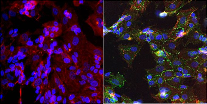
Immunofluorescent analysis of N-Cadherin in SHSY5Y cells. Formalin-fixed cells were permeabilized with 0.1% Triton X-100 in TBS for 10 minutes at room temperature. Cells were blocked with 1% BSA for 15 minutes at room temperature. Cells were probed without (left panel) or with (right panel) N-Cadherin antibody [8C11] at a dilution of 1:100 for at least 1 hour at room temperature, washed with PBS, and incubated with a proper secondary antibody. F-Actin (red) was stained with Phalloidin and nuclei (blue) were stained with Hoechst 33342 dye. Images were taken at 20X magnification.Application
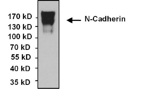
Immunoprecipitation of N-Cadherin was performed using SHSY5Y whole cell lysate. Antigen-antibody complexes were formed by incubating 300ug of lysate with 5ug of N-Cadherin antibody [8C11] overnight on a rocking platform at 4°C. The immune complexes were captured on 50ul Protein A/G Agarose , washed extensively, and eluted. The sample was resolved on a 4-20% Tris-HCl polyacrylamide gel, transferred to a PVDF membrane, and blocked with 5% BSA/TBS-0.1%Tween for at least 1 hour. The membrane was probed with N-Cadherin antibody [8C11] at a dilution of 1:1000 overnight rotating at 4°C, washed in TBST, and probed with an proper secondary antibody. Chemiluminescent detection was performed.Application
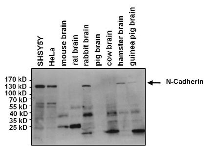
Western blot analysis of N-Cadherin in 25 ug of various lysates. Proteins were transferred to a PVDF membrane and blocked with 5% BSA/TBST for at least 1 hour. The membrane was probed with N-Cadherin antibody [8C11] at a dilution of 1:1000 overnight at 4°C on a rocking platform, washed in TBS-0.1%Tween-20, and probed with a HRP-conjugated secondary antibody for 1 hour. Chemiluminescent detection was performed. Note: analysis indicates both reactive and non-reactive species for N-cadherin.Application
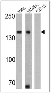
WB analysis of HeLa, HUVEC and C2C12 lysates (25 ug per lane) using N-Cadherin antibody [8C11] at a dilution of 1:1000 (HeLa) and 1:500 (HUVEC and C2C12).Application
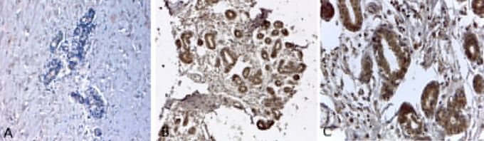
IHC-P analysis of human breast tissues with an isotype control (A) or N-Cadherin antibody [8C11] (B and C) at a concentration of 2ug/ml. (C) shows the magnified section of (B). To expose target proteins, antigen retrieval was performed using HEIR with a buffer (pH 6.2).| Product Name | N-Cadherin antibody |
|---|---|
| Antibody Type | Primary Antibodies |
| Product description | Mouse monoclonal antibody [8C11] to N-CadherinThis gene is a classical cadherin from the cadherin superfamily. The encoded protein is a calcium dependent cell-cell adhesion glycoprotein comprised of five extracellular cadherin repeats, a transmembrane region and a highly conserved cytoplasmic tail. The protein functions during gastrulation and is required for establishment of left-right asymmetry. At certain central nervous system synapses, presynaptic to postsynaptic adhesion is mediated at least in part by this gene product. [provided by RefSeq, Jul 2008] |
| Immunogen | Bacterial fusion protein corresponding to the extracellular domain of N-cadherin. |
| Conjugation | Agarose |
| Modification | Acetylated |
| Clonality | Monoclonal |
|---|---|
| Isotype | IgG1 |
| Clone Number | 8C11 |
| Host Species | Mouse |
| Tested Applications | ICC/IFIHC-PIPWB |
| WB:1:500~1:2000 IHC-P:1:50~1 ICC/IF:1:100~1:500 |
|
| Species Reactivity | AvianHamsterHumanRabbit |
| Concentration | 1mg/ml (Please refer to the vial label for the specific concentration) |
| Purification | Protein G |
| Gene Synonyms | CDw325 CD325 CDHN cadherin 2 type 1 N-cadherin (neuronal) NCAD CDH2 |
|---|---|
| Target | CD325/N Cadherin/CDH2 |
Application
Immunofluorescent analysis of N-Cadherin in SHSY5Y cells. Formalin-fixed cells were permeabilized with 0.1% Triton X-100 in TBS for 10 minutes at room temperature. Cells were blocked with 1% BSA for 15 minutes at room temperature. Cells were probed without (left panel) or with (right panel) N-Cadherin antibody [8C11] at a dilution of 1:100 for at least 1 hour at room temperature, washed with PBS, and incubated with a proper secondary antibody. F-Actin (red) was stained with Phalloidin and nuclei (blue) were stained with Hoechst 33342 dye. Images were taken at 20X magnification.
Application
Immunoprecipitation of N-Cadherin was performed using SHSY5Y whole cell lysate. Antigen-antibody complexes were formed by incubating 300ug of lysate with 5ug of N-Cadherin antibody [8C11] overnight on a rocking platform at 4°C. The immune complexes were captured on 50ul Protein A/G Agarose , washed extensively, and eluted. The sample was resolved on a 4-20% Tris-HCl polyacrylamide gel, transferred to a PVDF membrane, and blocked with 5% BSA/TBS-0.1%Tween for at least 1 hour. The membrane was probed with N-Cadherin antibody [8C11] at a dilution of 1:1000 overnight rotating at 4°C, washed in TBST, and probed with an proper secondary antibody. Chemiluminescent detection was performed.
Application
Western blot analysis of N-Cadherin in 25 ug of various lysates. Proteins were transferred to a PVDF membrane and blocked with 5% BSA/TBST for at least 1 hour. The membrane was probed with N-Cadherin antibody [8C11] at a dilution of 1:1000 overnight at 4°C on a rocking platform, washed in TBS-0.1%Tween-20, and probed with a HRP-conjugated secondary antibody for 1 hour. Chemiluminescent detection was performed. Note: analysis indicates both reactive and non-reactive species for N-cadherin.
Application
WB analysis of HeLa, HUVEC and C2C12 lysates (25 ug per lane) using N-Cadherin antibody [8C11] at a dilution of 1:1000 (HeLa) and 1:500 (HUVEC and C2C12).
Application
IHC-P analysis of human breast tissues with an isotype control (A) or N-Cadherin antibody [8C11] (B and C) at a concentration of 2ug/ml. (C) shows the magnified section of (B). To expose target proteins, antigen retrieval was performed using HEIR with a buffer (pH 6.2).| Application Notes | WB:1:500~1:2000 IHC-P:1:50~1 ICC/IF:1:100~1:500 |
|---|
| Form | Liquid |
|---|---|
| Storage Instructions | -20° C, Avoid Freeze/Thaw Cycles |
| Storage Buffer | PBS containing 0.05 % sodium azide and 1 mg/ml BSA |


