Application
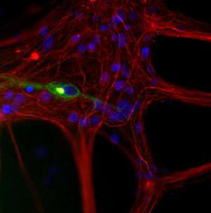
Immunocytochemistry/Immunofluorescence: 68kDa Neurofilament Light Antibody - Mixed neuron/glia cultures from newborn rat brain stained antibody to peripherin (green) and rabbit polyclonal antibody to NF-L RPCA-NF-L (red channel). A class of large neurons, like the one in the middle of this image, contain peripherin, while the majority of neurons and their processes contain NF-L and not peripherin. Interestingly, the periperin positive cells often contain a cytoplasmic inclusion next to the nucleus which stains for both peripherin and NF-L, and so appears golden in this kind of image. The blue channel reveals the localization of DNAApplication
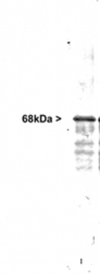
Western Blot: Neurofilament light chain Antibody - Western blot of whole rat cerebellum homogenate stained with , at dilution of 1:20,000. A prominent band running with an apparent SDS-PAGE molecular weight of ~68kDa corresponds to rodent NF-L. Human, cow and bovine NF-L run a little slower, at about 70kDa.| Product Name | NF-L antibody |
|---|---|
| Antibody Type | Primary Antibodies |
| Product description | Rabbit Polyclonal antibody to NF-LNeurofilaments are the 10nm or intermediate filament proteins found specifically in neurons, and are composed predominantly of three major proteins called NF-L, NF-M and NF-H. NF-L is the neurofilament light or low molecular weight polypeptide and runs on SDS-PAGE gels at about 68kDa. Antibodies to NF-L are useful for identifying neuronal cells and their processes in tissue sections and in tissue culture. NF-L antibody can also be useful in the diagnostics of neurofilament accumulations seen in many neurological diseases, such as Lou Gehrig's disease or Alzheimer's disease. |
| Immunogen | Native NF-L purified from pig spinal cord |
| Clonality | Polyclonal |
|---|---|
| Host Species | Rabbit |
| Tested Applications | ICC/IFIHC-FIHC-PWB |
| For IHC-Fr: Use at a dilution of 1:1,000. For IHC-P: Use at a dilution of 1:1,000. For WB: Use at a dilution of 1:5,000. Optimal dilutions/concentrations should be determined by the end user.: |
|
| Species Reactivity | HumanMousePigRatMammalian speciesBovine |
| Concentration | 1mg/ml |
| Purification | Affinity purified |
| Gene Synonyms | NF68 NEFL P07196 CMT2E NFL CMT1F NF-L 4747 162280 NF L |
|---|---|
| Target | NF-L |
| Tissue Specificity | Recognizes the light neurofilament subunit (~68 kDa). |
Application
Immunocytochemistry/Immunofluorescence: 68kDa Neurofilament Light Antibody - Mixed neuron/glia cultures from newborn rat brain stained antibody to peripherin (green) and rabbit polyclonal antibody to NF-L RPCA-NF-L (red channel). A class of large neurons, like the one in the middle of this image, contain peripherin, while the majority of neurons and their processes contain NF-L and not peripherin. Interestingly, the periperin positive cells often contain a cytoplasmic inclusion next to the nucleus which stains for both peripherin and NF-L, and so appears golden in this kind of image. The blue channel reveals the localization of DNA
Application
Western Blot: Neurofilament light chain Antibody - Western blot of whole rat cerebellum homogenate stained with , at dilution of 1:20,000. A prominent band running with an apparent SDS-PAGE molecular weight of ~68kDa corresponds to rodent NF-L. Human, cow and bovine NF-L run a little slower, at about 70kDa.| Positive Control | Rat cerebellum |
|---|---|
| Application Notes | For IHC-Fr: Use at a dilution of 1:1,000. For IHC-P: Use at a dilution of 1:1,000. For WB: Use at a dilution of 1:5,000. Optimal dilutions/concentrations should be determined by the end user.: |
| Form | Liquid |
|---|---|
| Storage Instructions | Keep as concentrated solution. Store at 4ºC short term. For extended storage aliquot and store at -20ºC or below. Avoid freeze-thaw cycles. |
| Storage Buffer | Whole antiserum containing no preservative |


