Application
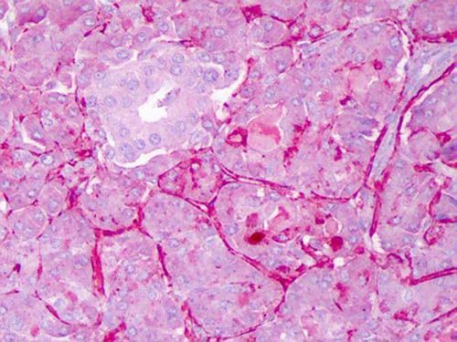
IHC-P analysis of exocrine glands of human pancreas using Rabbit anti-Notch1 antibody at a dilution of 1:200. Staining: moderate to strong membranous staining and faint to moderate cytoplasmic staining. Islets showed faint staining.Application
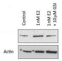
Western Blot of Rabbit anti-Notch1 antibody . Lane 1: MCF-7 control lysate. Lane 2: MCF-7 +1 nM 17ß-estradiol. Lane 3: MCF-7 + 10 µM gamma secretase inhibitor. Load: 35 µg per lane. Primary antibody: Notch1 antibody at 1:500 for overnight at 4°C. Rabbit secondary antibody at 1:10,000 for 45 min at RT.Predicted/Observed size: 80 kDa for Notch1.Application
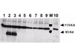
Rabbit anti-Human NOTCH 1 (Cleaved N Terminal) was used at a 1:500 dilution to detect mouse Notch 1 by Western blot in 293 cells transiently transfected with myc-tagged mouse Notch. Bound antibody was detected with a 1:10,000 dilution of HRP conjugated Goat anti Rabbit IgG, and color development. Lane M: markers. Lane 1: No transfection. Lane 2: N1 (mouse deleted extracellular domain)-myc. Lane 3: N1 (mouse intracellular domain)-myc. Lane 4: N2 (mouse deleted extracellular domain)-myc. Lane 5: N2 (mouse intracellular domain)-myc. Lane 6: N3 (mouse deleted extracellular domain)-myc. Lane 7: N3 (mouse intracellular domain)-myc. Lane 8: N4 (mouse deleted extracellular domain)-myc. Lane 9: N4 (mouse intracellular domain)-myc. Lane 10: N1 (mouse deleted extracellular domain)(V to G)-myc.| Product Name | NOTCH1 antibody |
|---|---|
| Antibody Type | Primary Antibodies |
| Product description | Rabbit Polyclonal antibody to NOTCH1Notch 1 is synthesized in the endoplasmic reticulum as an inactive form, which is proteolytically cleaved by a furin-like convertase (S1 cleavage) in the trans-golgi network before it reaches the plasma membrane to yield an active, ligand-accessible form. Cleavage results in a C-terminal fragment N(TM) and a N-terminal fragment N(EC). Following ligand binding, it is cleaved (S2 cleavage) by TNF-alpha converting enzyme (TACE) to yield a membrane-associated intermediate fragment called Notch extracellular truncation (NEXT). This fragment is then cleaved by presenilin-dependent gamma-secretase (S3 cleavage) to release the intracellular domain (NICD) from the membrane. |
| Immunogen | Peptide: VLLSRKRRRQHGQC (aa1755-1767).This is the N-terminal sequence of the cleaved N intracellular domain (NICD). |
| Clonality | Polyclonal |
|---|---|
| Host Species | Rabbit |
| Tested Applications | ELISAICC/IFIHC-PNeutWB |
| *Optimal dilutions/concentrations should be determined by the researcher. | |
| Species Reactivity | HumanMouseRat |
| Concentration | 1mg/ml |
| Purification | Affinity purified |
| Gene Synonyms | HN1 , TAN1 |
|---|---|
| Target | Notch1 |
Application
IHC-P analysis of exocrine glands of human pancreas using Rabbit anti-Notch1 antibody at a dilution of 1:200. Staining: moderate to strong membranous staining and faint to moderate cytoplasmic staining. Islets showed faint staining.
Application
Western Blot of Rabbit anti-Notch1 antibody . Lane 1: MCF-7 control lysate. Lane 2: MCF-7 +1 nM 17ß-estradiol. Lane 3: MCF-7 + 10 µM gamma secretase inhibitor. Load: 35 µg per lane. Primary antibody: Notch1 antibody at 1:500 for overnight at 4°C. Rabbit secondary antibody at 1:10,000 for 45 min at RT.Predicted/Observed size: 80 kDa for Notch1.
Application
Rabbit anti-Human NOTCH 1 (Cleaved N Terminal) was used at a 1:500 dilution to detect mouse Notch 1 by Western blot in 293 cells transiently transfected with myc-tagged mouse Notch. Bound antibody was detected with a 1:10,000 dilution of HRP conjugated Goat anti Rabbit IgG, and color development. Lane M: markers. Lane 1: No transfection. Lane 2: N1 (mouse deleted extracellular domain)-myc. Lane 3: N1 (mouse intracellular domain)-myc. Lane 4: N2 (mouse deleted extracellular domain)-myc. Lane 5: N2 (mouse intracellular domain)-myc. Lane 6: N3 (mouse deleted extracellular domain)-myc. Lane 7: N3 (mouse intracellular domain)-myc. Lane 8: N4 (mouse deleted extracellular domain)-myc. Lane 9: N4 (mouse intracellular domain)-myc. Lane 10: N1 (mouse deleted extracellular domain)(V to G)-myc.| Application Notes | *Optimal dilutions/concentrations should be determined by the researcher. |
|---|
| Form | Liquid |
|---|---|
| Storage Instructions | Store vial at -20° C prior to opening. Aliquot contents and freeze at -20° C or below for extended storage. Avoid cycles of freezing and thawing. |
| Storage Buffer | 0.02 M Potassium Phosphate, 0.15 M Sodium Chloride, pH 7.2 and 0.1% (w/v) Sodium Azide |


