Application
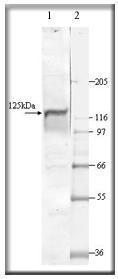
at a 1:500 dilution, staining PGCA by Western blot. Lane 1: PC-PGCA; Lane 2: MWM.| Product Name | NPR-A antibody |
|---|---|
| Antibody Type | Primary Antibodies |
| Product description | Rabbit polyclonal to NPR-ACyclic GMP (cGMP) is a key messenger in several signal transduction pathways. The intracellular levels of cGMP are maintained by the activity of opposing enzymes: synthesizing gualylyl cyclases (GC) and hydrolyzing phosphodiesterases (PDEs). The synthesizing enzymes (GCs) are found in two forms: cytosolic (soluble) and membrane bound (particulate). In the last several years, new members of particulate guanylate cyclase family have been identified, and significant progress has been made in understanding the molecular mechanisms that underlie the regulation of this family of enzymes. In response to G protein coupled receptor stimulation, the cGMP can be produced from GTP by either cytoplasmic, soluble guanylate cyclase (sGC) that are heterodimers stimulated by nitric oxide and carbon monoxide, or by particulate membrane bound guanylyl cyclases which are activated in a complex mechanism by natriuretic peptides. Particulate GC (PGCs) have 7 different isoforms, PGC A through PGC G, and are expressed in most tissues in isoform specific manner. There is significant structural homology among various PGCs. There is a large N terminal extracellular domain (ECD), a single TMD, and a large intracellular domain with protein kinase activity (KLD), a C terminal catalytic domain (CD), and in between is a dimmerization domain (DD). Both PGC A and PGC B are posphorylated at Serine residues in the KLD. Non ionic detergents stimulated particulate guanylate cyclase activity in cerebral cortex of rat 8- to 12-fold, while stimulation of soluble enzyme was 1.3- to 2.5-fold. It has been shown that a significant number of hippcampal astrocytes (67%) contained both soluble and particulate guanylate cyclases in the same cell. |
| Immunogen | Synthetic peptide: CVRTYWLLGERGCSTRG (C terminal). |
| Clonality | Polyclonal |
|---|---|
| Isotype | IgG |
| Host Species | Rabbit |
| Tested Applications | WB |
| WB: Use at a dilution of 1/500. Detects a band of approximately 127 kDa. Optimal dilutions/concentrations should be determined by the end user.: |
|
| Species Reactivity | HumanMouseRat |
| Concentration | 1mg/ml |
| Purification | Affinity purified |
| Gene Synonyms | Particulate Guanylate Cyclase A |
|---|---|
| Target | PGCA |
| Tissue Specificity | The selective antibodies were generated against conserved sequences near the N terminus and the C terminal end of the protein that are unique to PGCA protein. The PGCA antibody labels a 127 to 130 kDa protein in various tissues. |
| Cellular Localization | Type I membrane protein. |
Application
at a 1:500 dilution, staining PGCA by Western blot. Lane 1: PC-PGCA; Lane 2: MWM.| Positive Control | Brain, kidney and spleen |
|---|---|
| Application Notes | WB: Use at a dilution of 1/500. Detects a band of approximately 127 kDa. Optimal dilutions/concentrations should be determined by the end user.: |
| Form | Liquid |
|---|---|
| Storage Instructions | Keep as concentrated solution. Store at 4ºC short term. For extended storage aliquot and store at -20ºC or below. Avoid freeze-thaw cycles. |
| Storage Buffer | phosphate buffered saline , pH 7.4, 150mM NaCl, 0.02% sodium azide and 50% glycerol. |


