Application
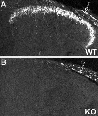
Image of NPY Y2 receptor immunoreactivity in mouse dorsal root ganglion of wild type (A) and NPY Y2 receptor Knockout (B) animals. Note that unspecific staining is observed in thick, possibly myelinated fibers. Image courtesy of Dr. Tomas Hkfelt, Karolins| Product Name | NPY Y2 antibody |
|---|---|
| Antibody Type | Primary Antibodies |
| Product description | Rabbit polyclonal NPY Y2 Receptor antiserumThis NPY Y2 antibody has been extensively characterized at the spinal level and is currently being characerized in brain.? The antibody has been tested in both rat and mouse and appears to work better on mose tissue, although positive staining is seen in rat.? For full characterizarion in dorsal root ganglion and spinal cord see: J Comp Neurol. 2005 Aug 29;489(3):328-48. |
| Clonality | Polyclonal |
|---|---|
| Host Species | Rabbit |
| Tested Applications | IHC |
| Immunohistochemistry 1:500 -1:1000. Dilutions listed only as a recommendation. Optimal dilution should be determined by investigator. Immunohistochemistry: Antiserum was used on perfusion fixed tissue. Perfusion: 1) calcium-free Tyrode’s solution, 2) paraformaldehyde-picric acid fixative, and 3) 10% sucrose in PBS as a cryo-protectant. Desired tissues were dissected and stored overnight in 10% sucrose in PBS. Slide-mounted tissue sections were processed for indirect immunofluorescence. Slides were incubated with blocking buffer for 1 hour at room temperature. Primary antiserum was diluted with blocking buffer to the appropriate working concentration. Blocking buffer was removed and slides were incubated for 18-24 hours at 4oC with primary antiserum. Slides were rinsed 3 times and then incubated with secondary antibodies for 1 hour at room temperature. Slides were again rinsed 3 times and coverslipped. Staining was examined using fluorescence microscopy.: |
|
| Species Reactivity | MouseRat |
| Concentration | 1mg/ml |
| Target | NPY Y2 |
|---|
Application
Image of NPY Y2 receptor immunoreactivity in mouse dorsal root ganglion of wild type (A) and NPY Y2 receptor Knockout (B) animals. Note that unspecific staining is observed in thick, possibly myelinated fibers. Image courtesy of Dr. Tomas Hkfelt, Karolins| Application Notes | Immunohistochemistry 1:500 -1:1000. Dilutions listed only as a recommendation. Optimal dilution should be determined by investigator. Immunohistochemistry: Antiserum was used on perfusion fixed tissue. Perfusion: 1) calcium-free Tyrode’s solution, 2) paraformaldehyde-picric acid fixative, and 3) 10% sucrose in PBS as a cryo-protectant. Desired tissues were dissected and stored overnight in 10% sucrose in PBS. Slide-mounted tissue sections were processed for indirect immunofluorescence. Slides were incubated with blocking buffer for 1 hour at room temperature. Primary antiserum was diluted with blocking buffer to the appropriate working concentration. Blocking buffer was removed and slides were incubated for 18-24 hours at 4oC with primary antiserum. Slides were rinsed 3 times and then incubated with secondary antibodies for 1 hour at room temperature. Slides were again rinsed 3 times and coverslipped. Staining was examined using fluorescence microscopy.: |
|---|
| Form | Liquid |
|---|---|
| Storage Instructions | Upon Receipt - Keep as concentrated solution. Aliquot and store at -20ºC or below. Avoid freeze-thaw cycles. |
| Storage Buffer | 1.28 mg/ml In PBS with 0.05% sodium Azide |


