Application
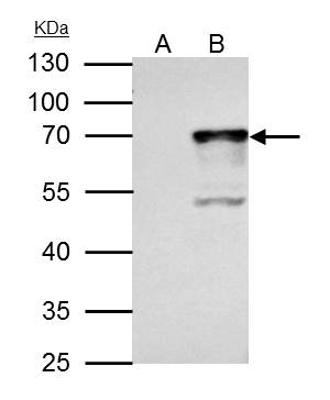
NRF1 antibody immunoprecipitates NRF1 protein in IP experiments. IP samples: 293T whole cell extract A. Control with 4 μg of preimmune Rabbit IgG B. Immunoprecipitation of NRF1 protein by 4 μg NRF1 antibody 10 % SDS-PAGE The immunoprecipitated NRF1 protein was detected by NRF1 antibody diluted at 1:1000. [EasyBlot anti-rabbit IgG was used as a secondary reagent]Application
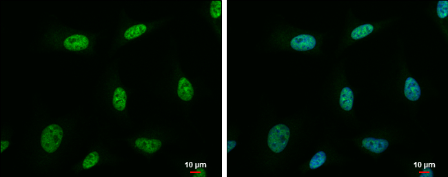
NRF1 antibody detects NRF1 protein at nucleus by immunofluorescent analysis. Sample: HeLa cells were fixed in 4% paraformaldehyde at RT for 15 min. Green: NRF1 protein stained by NRF1 antibody diluted at 1:500. Blue: Hoechst 33342 staining. Scale bar = 10 μm.Application
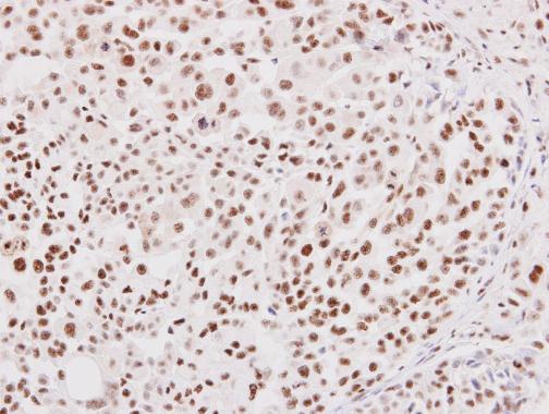
NRF1 antibody detects NRF1 protein at nucleus on human lung adenocarcinoma by immunohistochemical analysis. Sample: Paraffin-embedded human lung adenocarcinoma. NRF1 antibody diluted at 1:250.Application
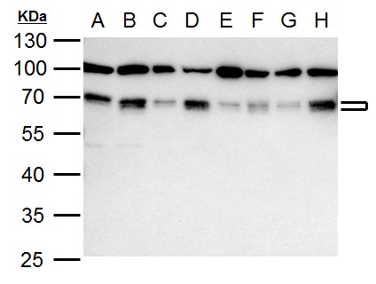
NRF1 antibody detects NRF1 protein by western blot analysis. A. 30 μg 293T whole cell lysate/extract B. 30 μg 293T nuclear lysate/extract C. 30 μg A431 whole cell lysate/extract | D. 30 μg A431 nuclear lysate/extract E. 30 μg HeLa whole cell lysate/extract F. 30 μg HeLa nuclear lysate/extract G. 30 μg HepG2 whole cell lysate/extract H. 30 μg HepG2 nuclear lysate/extract 10 % SDS-PAGE NRF1 antibody dilution: 1:1000Application
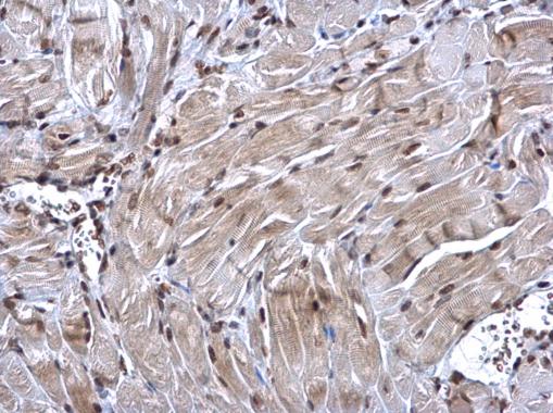
NRF1 antibody detects NRF1 protein at nucleus on mouse muscle by immunohistochemical analysis. Sample: Paraffin-embedded mouse muscle. NRF1 antibody dilution: 1:500.Application
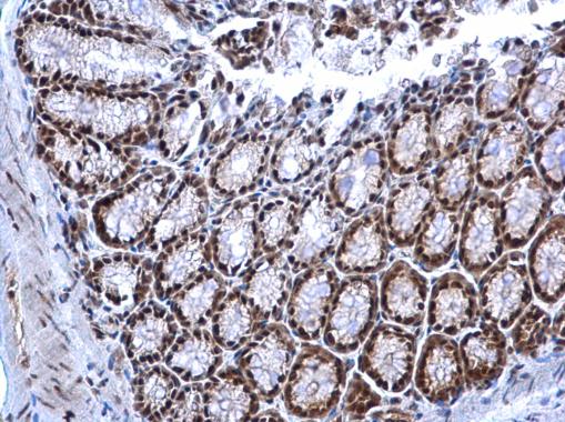
NRF1 antibody detects NRF1 protein at nucleus on mouse colon by immunohistochemical analysis. Sample: Paraffin-embedded mouse colon. NRF1 antibody dilution: 1:500.| Product Name | NRF1 antibody |
|---|---|
| Antibody Type | Primary Antibodies |
| Product description | Rabbit Polyclonal antibody to NRF1 (nuclear respiratory factor 1)This gene encodes a protein that homodimerizes and functions as a transcription factor which activates the expression of some key metabolic genes regulating cellular growth and nuclear genes required for respiration, heme biosynthesis, and mitochondrial DNA transcription and replication. The protein has also been associated with the regulation of neurite outgrowth. Alternate transcriptional splice variants, which encode the same protein, have been characterized. Additional variants encoding different protein isoforms have been described but they have not been fully characterized. Confusion has occurred in bibliographic databases due to the shared symbol of NRF1 for this gene and for "nuclear factor (erythroid-derived 2)-like 1" which has an official symbol of NFE2L1. [provided by RefSeq] |
| Immunogen | Recombinant protein encompassing a sequence within the center region of human NRF1. The exact sequence is proprietary. |
| Clonality | Polyclonal |
|---|---|
| Isotype | IgG |
| Host Species | Rabbit |
| Tested Applications | ICC/IFIHC-PIPWB |
| *Optimal dilutions/concentrations should be determined by the researcher.
: |
|
| Species Reactivity | HumanMouse |
| Predicted Reactivity | Rat, Zebrafish, Xenopus laevis, Chicken, BovineRat (100%), Zebrafish (92%), Xenopus laevis (93%), Chicken (99%), Bovine (100%) |
| Concentration | 1mg/ml (Please refer to the vial label for the specific concentration) |
| Purification | Affinity purified |
| Gene Synonyms | ALPHA-PAL antibody NRF1 antibody NRF-1 antibody nuclear respiratory factor 1 antibody alpha palindromic-binding protein antibody |
|---|---|
| Molecular Weight(MW) | 54 kDa (note) (3) |
| Cellular Localization | Nucleus |
Application
NRF1 antibody immunoprecipitates NRF1 protein in IP experiments. IP samples: 293T whole cell extract A. Control with 4 μg of preimmune Rabbit IgG B. Immunoprecipitation of NRF1 protein by 4 μg NRF1 antibody 10 % SDS-PAGE The immunoprecipitated NRF1 protein was detected by NRF1 antibody diluted at 1:1000. [EasyBlot anti-rabbit IgG was used as a secondary reagent]
Application
NRF1 antibody detects NRF1 protein at nucleus by immunofluorescent analysis. Sample: HeLa cells were fixed in 4% paraformaldehyde at RT for 15 min. Green: NRF1 protein stained by NRF1 antibody diluted at 1:500. Blue: Hoechst 33342 staining. Scale bar = 10 μm.
Application
NRF1 antibody detects NRF1 protein at nucleus on human lung adenocarcinoma by immunohistochemical analysis. Sample: Paraffin-embedded human lung adenocarcinoma. NRF1 antibody diluted at 1:250.
Application
NRF1 antibody detects NRF1 protein by western blot analysis. A. 30 μg 293T whole cell lysate/extract B. 30 μg 293T nuclear lysate/extract C. 30 μg A431 whole cell lysate/extract | D. 30 μg A431 nuclear lysate/extract E. 30 μg HeLa whole cell lysate/extract F. 30 μg HeLa nuclear lysate/extract G. 30 μg HepG2 whole cell lysate/extract H. 30 μg HepG2 nuclear lysate/extract 10 % SDS-PAGE NRF1 antibody dilution: 1:1000
Application
NRF1 antibody detects NRF1 protein at nucleus on mouse muscle by immunohistochemical analysis. Sample: Paraffin-embedded mouse muscle. NRF1 antibody dilution: 1:500.
Application
NRF1 antibody detects NRF1 protein at nucleus on mouse colon by immunohistochemical analysis. Sample: Paraffin-embedded mouse colon. NRF1 antibody dilution: 1:500.| Positive Control | 293T , 293T nuclear , A431 , A431 nuclear , HeLa , HeLa nuclear , HepG2 , HepG2 nuclear |
|---|---|
| Application Notes | *Optimal dilutions/concentrations should be determined by the researcher.
: |
| Form | Liquid |
|---|---|
| Storage Instructions | Keep as concentrated solution. Aliquot and store at -20ºC or below. Avoid multiple freeze-thaw cycles. |
| Storage Buffer | 1XPBS, 20% Glycerol (pH7). 0.025% ProClin 300 was added as a preservative. |


