Application
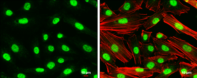
Nurr1 antibody detects Nurr1 protein at nucleus by immunofluorescent analysis. Sample: SK-N-SH cells were fixed in 4% paraformaldehyde at RT for 15 min. Green: Nurr1 protein stained by Nurr1 antibody diluted at 1:500. Red: Phalloidin, a cytoskeleton marker, diluted at 1:50. Scale bar = 10 μm.Application
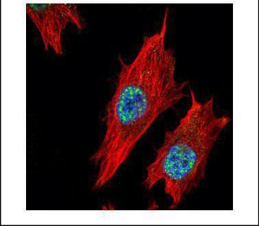
Confocal immunofluorescence analysis (Olympus FV10i) of paraformaldehyde-fixed HeLa, using Nurr1 antibody (Green) at 1:500 dilution. Alpha-tubulin filaments were labeled with (Red) at 1:2000.| Product Name | Nurr1 antibody |
|---|---|
| Antibody Type | Primary Antibodies |
| Product description | Rabbit Polyclonal antibody to Nurr1 (nuclear receptor subfamily 4, group A, member 2)This gene encodes a member of the steroid-thyroid hormone-retinoid receptor superfamily. The encoded protein may act as a transcription factor. Mutations in this gene have been associated with disorders related to dopaminergic dysfunction, including Parkinson disease, schizophernia, and manic depression. Misregulation of this gene may be associated with rheumatoid arthritis. Alternatively spliced transcript variants have been described, but their biological validity has not been determined. [provided by RefSeq] |
| Immunogen | Recombinant protein encompassing a sequence within the center region of human Nurr1. The exact sequence is proprietary. |
| Clonality | Polyclonal |
|---|---|
| Isotype | IgG |
| Host Species | Rabbit |
| Tested Applications | ICC/IF |
| *Optimal dilutions/concentrations should be determined by the researcher.
: |
|
| Species Reactivity | Human |
| Predicted Reactivity | Mouse, Rat, Pig, Rhesus Monkey, BovineMouse (100%), Rat (100%), Pig (100%), Rhesus Monkey (100%), Bovine (99%) |
| Concentration | 1mg/ml |
| Purification | Affinity purified |
| Gene Synonyms | HZF-3 antibody NOT antibody NURR1 antibody RNR1 antibody TINUR antibody NR4A2 antibody orphan nuclear receptor NR4A2 antibody transcriptionally inducible nuclear receptor related 1 antibody nuclear receptor related 1 antibody NGFI-B/nur77 beta-typ |
|---|---|
| Molecular Weight(MW) | 67 kDa (note) |
| Cellular Localization | Nucleus |
Application
Nurr1 antibody detects Nurr1 protein at nucleus by immunofluorescent analysis. Sample: SK-N-SH cells were fixed in 4% paraformaldehyde at RT for 15 min. Green: Nurr1 protein stained by Nurr1 antibody diluted at 1:500. Red: Phalloidin, a cytoskeleton marker, diluted at 1:50. Scale bar = 10 μm.
Application
Confocal immunofluorescence analysis (Olympus FV10i) of paraformaldehyde-fixed HeLa, using Nurr1 antibody (Green) at 1:500 dilution. Alpha-tubulin filaments were labeled with (Red) at 1:2000.| Positive Control | Target recombinant protein |
|---|---|
| Application Notes | *Optimal dilutions/concentrations should be determined by the researcher.
: |
| Form | Liquid |
|---|---|
| Storage Instructions | Keep as concentrated solution. Aliquot and store at -20ºC or below. Avoid multiple freeze-thaw cycles. |
| Storage Buffer | 0.1M Tris, 0.1M Glycine, 20% Glycerol (pH7). 0.01% Thimerosal was added as a preservative. |


