Application
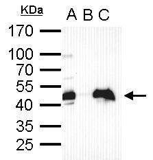
Oct4 antibody immunoprecipitates Oct4 protein in IP experiments. IP Sample: cell lysate/extract of Oct4 gene transfected 293T cells A. Cell lysate/extract of transfected 293T cell B. Control with 2 μg of preimmune rabbit IgG C. Immunoprecipitation of Oct4 by 2 μg of Oct4 antibody 12% SDS-PAGE The immunoprecipitated Oct4 protein was detected by Oct4 antibody diluted at 1:1000. EasyBlot anti-rabbit IgG was used as a secondary reagent.Application
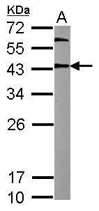
Sample (20 µg of whole cell lysate) A: mouse ESC 12% SDS PAGE diluted at 1:10000 The HRP-conjugated anti-rabbit IgG antibody was used to detect the primary antibody.Application
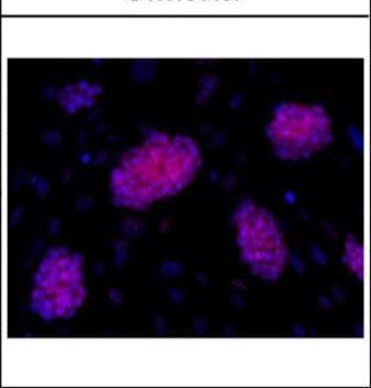
Immunofluorescence analysis of mESCs on CF1 feeders, using OCT3/4 antibody at 1:200 dilution.Application
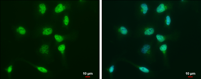
Oct4 antibody detects Oct4 protein at nucleus by immunofluorescent analysis. Sample: NT2D1 cells were fixed in 4% paraformaldehyde at RT for 15 min. Green: Oct4 protein stained by Oct4 antibody diluted at 1:500. Blue: Hoechst 33342 staining. Scale bar = 10 μm.Application
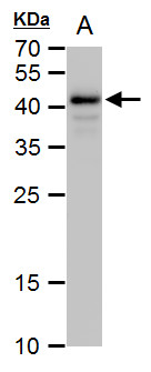
Oct4 antibody detects Oct4 protein by western blot analysis. A. 30 µg human ESC whole cell extract 12% SDS-PAGE Oct4 antibody dilution: 1:10000 The HRP-conjugated anti-rabbit IgG antibody was used to detect the primary antibody.Application
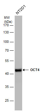
OCT4 antibody detects OCT4 protein by western blot analysis. Whole cell extracts (30 µg) was separated by 10% SDS-PAGE, and the membrane was blotted with OCT4 antibody at a dilution of 1:50000. The HRP-conjugated anti-rabbit IgG antibody was used to detect the primary antibody.Application
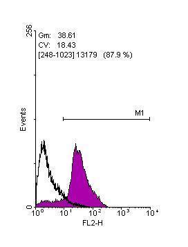
Flow cytometry on human embryonic stem cells, staining with Oct4 antibody at 1:100 dilution(purple) or rabbit IgG (black).Application
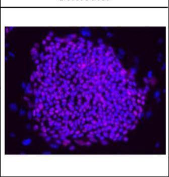
Immunofluorescence analysis of hESCs on CF1 feeders, using OCT3/4 antibody at 1:200 dilution.Application
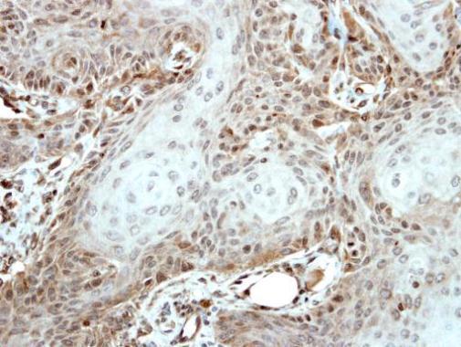
Immunohistochemical analysis of paraffin-embedded Cal27 xenograft, using OCT3/4 antibody at 1:500 dilution.| Product Name | Oct4 antibody |
|---|---|
| Antibody Type | Primary Antibodies |
| Product description | Rabbit Polyclonal antibody to OCT4 (POU class 5 homeobox 1)This gene encodes a transcription factor containing a POU homeodomain. This transcription factor plays a role in embryonic development, especially during early embryogenesis, and it is necessary for embryonic stem cell pluripotency. A translocation of this gene with the Ewing's sarcoma gene, t(6;22)(p21;q12), has been linked to tumor formation. Alternative splicing, as well as usage of alternative translation initiation codons, results in multiple isoforms, one of which initiates at a non-AUG (CUG) start codon. Related pseudogenes have been identified on chromosomes 1, 3, 8, 10, and 12. [provided by RefSeq] |
| Immunogen | Recombinant protein encompassing a sequence within the center region of human Oct4. The exact sequence is proprietary. |
| Clonality | Polyclonal |
|---|---|
| Isotype | IgG |
| Host Species | Rabbit |
| Tested Applications | FACSICC/IFIHC-FIHC-PIPWB |
| WB:1:500~1:1000 ICC/IF:1:100~1:500 IHC:1:50~1:100 *Optimal dilutions/concentrations should be determined by the researcher.: |
|
| Species Reactivity | HumanMouse |
| Predicted Reactivity | Rat, Cat, Pig, Rabbit, Rhesus Monkey, Chimpanzee, BovineRat (83%), Cat (94%), Pig (93%), Rabbit (90%), Rhesus Monkey (98%), Chimpanzee (100%), Bovine (91%) |
| Concentration | 1mg/ml (Please refer to the vial label for the specific concentration) |
| Purification | Affinity purified |
| Gene Synonyms | MGC22487 antibody OTF-3 antibody OTF3 antibody OTF4 antibody POU5F1 antibody POU-type homeodomain-containing DNA-binding protein antibody octamer-binding protein 3 antibody POU domain transcription factor OCT4 antibody octamer-binding protein 4 ant |
|---|---|
| Molecular Weight(MW) | 39 kDa (note) |
| Cellular Localization | Nucleus |
Application
Oct4 antibody immunoprecipitates Oct4 protein in IP experiments. IP Sample: cell lysate/extract of Oct4 gene transfected 293T cells A. Cell lysate/extract of transfected 293T cell B. Control with 2 μg of preimmune rabbit IgG C. Immunoprecipitation of Oct4 by 2 μg of Oct4 antibody 12% SDS-PAGE The immunoprecipitated Oct4 protein was detected by Oct4 antibody diluted at 1:1000. EasyBlot anti-rabbit IgG was used as a secondary reagent.
Application
Sample (20 µg of whole cell lysate) A: mouse ESC 12% SDS PAGE diluted at 1:10000 The HRP-conjugated anti-rabbit IgG antibody was used to detect the primary antibody.
Application
Immunofluorescence analysis of mESCs on CF1 feeders, using OCT3/4 antibody at 1:200 dilution.
Application
Oct4 antibody detects Oct4 protein at nucleus by immunofluorescent analysis. Sample: NT2D1 cells were fixed in 4% paraformaldehyde at RT for 15 min. Green: Oct4 protein stained by Oct4 antibody diluted at 1:500. Blue: Hoechst 33342 staining. Scale bar = 10 μm.
Application
Oct4 antibody detects Oct4 protein by western blot analysis. A. 30 µg human ESC whole cell extract 12% SDS-PAGE Oct4 antibody dilution: 1:10000 The HRP-conjugated anti-rabbit IgG antibody was used to detect the primary antibody.
Application
OCT4 antibody detects OCT4 protein by western blot analysis. Whole cell extracts (30 µg) was separated by 10% SDS-PAGE, and the membrane was blotted with OCT4 antibody at a dilution of 1:50000. The HRP-conjugated anti-rabbit IgG antibody was used to detect the primary antibody.
Application
Flow cytometry on human embryonic stem cells, staining with Oct4 antibody at 1:100 dilution(purple) or rabbit IgG (black).
Application
Immunofluorescence analysis of hESCs on CF1 feeders, using OCT3/4 antibody at 1:200 dilution.
Application
Immunohistochemical analysis of paraffin-embedded Cal27 xenograft, using OCT3/4 antibody at 1:500 dilution.| Positive Control | hESC , mouse ESC , NT2D1 |
|---|---|
| Application Notes | WB:1:500~1:1000 ICC/IF:1:100~1:500 IHC:1:50~1:100 *Optimal dilutions/concentrations should be determined by the researcher.: |
| Form | Liquid |
|---|---|
| Storage Instructions | Keep as concentrated solution. Aliquot and store at -20ºC or below. Avoid multiple freeze-thaw cycles. |
| Storage Buffer | 1XPBS, 20% Glycerol (pH7). 0.025% ProClin 300 was added as a preservative. |


