Application
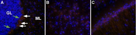
IHC analysis of rat (A) cerebellum, (B) parietal cortex and (C) hippocampus using Orexin Receptor 2 antibody at a dilution of 1:50 (red) and Hoechst 33342 (blue). GL: Golgi type neurons. ML: molecular layer.Application
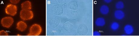
ICC/IF analysis of Colo-205 cells using Orexin Receptor 2 antibody at a dilution of 1:500 (red). (B) Live view of the same field as in (A). C. Nuclei were stained with Hoechst 33342 (blue).Application
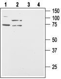
WB analysis of Colo-205 (lanes 1 and 3) and HT-29 (lanes 2 and 4) lysates using Orexin Receptor 2 antibody at a dilution of 1:200 in the absence (lens 1 and 2) or presence (lens 3 and 4) of blocking peptide.Application
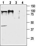
WB analysis of rat (lanes 1 and 3) and mouse (lanes 2 and 4) brain lysates using Orexin Receptor 2 antibody at a dilution of 1:400 in the absence (lens 1 and 2) or presence (lens 3 and 4) of blocking peptide.| Product Name | Orexin Receptor 2 antibody |
|---|---|
| Antibody Type | Primary Antibodies |
| Product description | Rabbit Polyclonal antibody to Orexin Receptor 2G-protein coupled receptor that binds orexin neuropeptides; involved in regulation of feeding behavior [RGD, Feb 2006] |
| Immunogen | Peptide (C)DRLARGRTSTESRKS, corresponding to amino acid residues 391-405 of rat OX2R (Accession P56719). Intracellular, C-terminus. |
| Clonality | Polyclonal |
|---|---|
| Host Species | Rabbit |
| Tested Applications | ICC/IFIHCWB |
| ICC/IF:1:200~1:500 IHC:1:50~1:200 WB:1:500~1:2000 Notes:Optimal dilutions/concentrations should be determined by the researcher. |
|
| Species Reactivity | HumanMouseRat |
| Concentration | 1mg/ml |
| Purification | Affinity purified |
| Gene Synonyms | Hcrtr2 hypocretin (orexin) receptor 2 |
|---|---|
| Target | Orexin Receptor 2 |
Application
IHC analysis of rat (A) cerebellum, (B) parietal cortex and (C) hippocampus using Orexin Receptor 2 antibody at a dilution of 1:50 (red) and Hoechst 33342 (blue). GL: Golgi type neurons. ML: molecular layer.
Application
ICC/IF analysis of Colo-205 cells using Orexin Receptor 2 antibody at a dilution of 1:500 (red). (B) Live view of the same field as in (A). C. Nuclei were stained with Hoechst 33342 (blue).
Application
WB analysis of Colo-205 (lanes 1 and 3) and HT-29 (lanes 2 and 4) lysates using Orexin Receptor 2 antibody at a dilution of 1:200 in the absence (lens 1 and 2) or presence (lens 3 and 4) of blocking peptide.
Application
WB analysis of rat (lanes 1 and 3) and mouse (lanes 2 and 4) brain lysates using Orexin Receptor 2 antibody at a dilution of 1:400 in the absence (lens 1 and 2) or presence (lens 3 and 4) of blocking peptide.| Application Notes | ICC/IF:1:200~1:500 IHC:1:50~1:200 WB:1:500~1:2000 Notes:Optimal dilutions/concentrations should be determined by the researcher. |
|---|
| Form | Liquid |
|---|---|
| Storage Instructions | Keep as concentrated solution. Aliquot and store at -20oC or below. Avoid multiple freeze-thaw cycles. |
| Storage Buffer | Reconstituted antibody containsA?phosphate buffered saline (PBS), pH 7.4, 1% BSA, 0.025% NaN3. |


