Application
ApplicationApplication
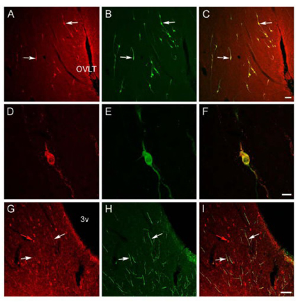
Confocal images showing OFQ (red), GnRH (green) and their colocalization (yellow) in the ovine preoptic area and hypothalamus. A-C: Low power view of the preoptic area at the level of the organum vasculosum of the lamina terminalis (OVLT) showing OFQ and GnRH cells, and their colocalization (e.g., white arrows). Bar = 100µm. D-F: High power images of a double-labeled OFQ/GnRH cell; note that the immunofluorescence for OFQ has a punctateappearance in the cytoplasm as opposed to the more diffuse appearance of the GnRH signal. Bar 26 = 20µm. G-I: Single-labeled OFQ cells (e.g., red arrow) in the arcuate nucleus. Note that many GnRH fibers coursing through this region are also OFQ-positive (e.g., white arrows). 3v = third ventricle. Bar = 100µm.| Product Name | Orphanin FQ antibody |
|---|---|
| Antibody Type | Primary Antibodies |
| Product description | Rabbit polyclonal Orphanin FQ antiserumOrphanin FQ (nociceptin, OFQ), a heptadecapeptide peptide, has been designated as an endogenous ligand for the ORL1 receptor. The amino acid sequence of OFQ has homology with other opioid peptides, especially the prodynorphin fragment ?dynorphin A, suggesting a close evolutionary relationship between the precursors. A diffuse distribution throughout the brain is seen with binding studies and in situ hybridization suggesting an extensive role for OFQ in a multitude of CNS functions. Immunocytochemical localizations in rat spinal cord have demonstrated OFQ abundance in superficial dorsal horn, lateral spinal nucleus and the region dorsal to the central canal. OFQ immunoreactivity was not affected by dorsal rhizotomy, indicating that in spinal cord the peptide is produced by central rather than primary afferent neurons. The localization is supported by its function in processing of nociceptive signals. |
| Immunogen | FGGFTGARKSARKLANQ Corresponding to residues 122-138 of rat Orphanin FQ |
| Clonality | Polyclonal |
|---|---|
| Host Species | Rabbit |
| Tested Applications | ICC/IFIHC |
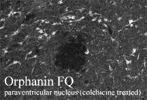
| Immunohistochemistry 1:500 (Dilutions listed only as a recommendation. Optimal dilution should be determined by investigator.) Immunohistochemistry: Antiserum was used on perfusion fixed tissue. Perfusion: 1) calcium-free Tyrode’s solution, 2) paraformaldehyde-picric acid fixative, and 3) 10% sucrose in PBS as a cryo-protectant. Desired tissues were dissected and stored overnight in 10% sucrose in PBS.Slide-mounted tissue sections were processed for indirect immunofluorescence. Slides were incubated with blocking buffer for 1 hour at room temperature. Primary antiserum was diluted with blocking buffer to the appropriate working concentration. Blocking buffer was removed and slides were incubated for 18-24 hours at 4oC with primary antiserum. Slides were rinsed 3 times and then incubated with secondary antibodies for 1 hour at room temperature. Slides were again rinsed 3 times and coverslipped. Staining was examined using fluorescence microscopy.: |
|
| Species Reactivity | RatPrimate |
| Concentration | 1mg/ml |
| Gene Synonyms | N23K Npnc1 |
|---|---|
| Target | Orphanin FQ |
Application
Application
Application
Confocal images showing OFQ (red), GnRH (green) and their colocalization (yellow) in the ovine preoptic area and hypothalamus. A-C: Low power view of the preoptic area at the level of the organum vasculosum of the lamina terminalis (OVLT) showing OFQ and GnRH cells, and their colocalization (e.g., white arrows). Bar = 100µm. D-F: High power images of a double-labeled OFQ/GnRH cell; note that the immunofluorescence for OFQ has a punctateappearance in the cytoplasm as opposed to the more diffuse appearance of the GnRH signal. Bar 26 = 20µm. G-I: Single-labeled OFQ cells (e.g., red arrow) in the arcuate nucleus. Note that many GnRH fibers coursing through this region are also OFQ-positive (e.g., white arrows). 3v = third ventricle. Bar = 100µm.| Application Notes | Immunohistochemistry 1:500 (Dilutions listed only as a recommendation. Optimal dilution should be determined by investigator.) Immunohistochemistry: Antiserum was used on perfusion fixed tissue. Perfusion: 1) calcium-free Tyrode’s solution, 2) paraformaldehyde-picric acid fixative, and 3) 10% sucrose in PBS as a cryo-protectant. Desired tissues were dissected and stored overnight in 10% sucrose in PBS.Slide-mounted tissue sections were processed for indirect immunofluorescence. Slides were incubated with blocking buffer for 1 hour at room temperature. Primary antiserum was diluted with blocking buffer to the appropriate working concentration. Blocking buffer was removed and slides were incubated for 18-24 hours at 4oC with primary antiserum. Slides were rinsed 3 times and then incubated with secondary antibodies for 1 hour at room temperature. Slides were again rinsed 3 times and coverslipped. Staining was examined using fluorescence microscopy.: |
|---|
| Form | Liquid |
|---|---|
| Storage Instructions | Upon Receipt - Keep as concentrated solution. Aliquot and store at -20ºC or below. Avoid freeze-thaw cycles. |
| Storage Buffer | Whole Serum |


