Application
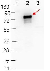
Western blot showing detection of 0.1 µg of recombinant OspB protein. Lane 1: Molecular weight markers. Lane 2: MBP-OspB fusion protein (arrow; expected MW = 72.7 kDa). Lane 3: MBP alone. Protein was run on a 4-20% gel, then transferred to 0.45 µm nitrocellulose. After blocking with 1% BSA-TTBS ) overnight at 4°C, primary antibody was used at 1:1000 at room temperature for 30 min. HRP-conjugated Goat-Anti-Rabbit secondary antibody was used at 1:40,000 in blocking buffer and imaged on the VersaDoc MP 4000 imaging system (Bio-Rad).| Product Name | OspB Antibody |
|---|---|
| Antibody Type | Primary Antibodies |
| Product description | Rabbit Polyclonal antibody to OspBOspB, is one of the major Outer Surface Proteins of the outer membrane of Borrelia burgdorferi, which is composed of various unique outer surface proteins (Osp) that have been characterized (OspA through OspF). The Osp proteins are lipoproteins anchored by N-terminally attached fatty acid molecules to the membrane. They are presumed to play a role in virulence, transmission, or survival in the tick. Two of the major surface Ag of Borrelia burgdorferi, the 31-kDa OspA and 34-kDa OspB proteins, show a high degree of sequence similarity, are encoded by a 49-kb plasmid and share a common promoter, and are coordinately transcribed. OspA, OspB, and OspD are expressed by B. burgdorferi residing in the gut of unfed ticks, suggesting that they promote the persistence of the spirochete in ticks between blood meals. OspB has a contributing role in the adherence of B. burgdorferi to the tick gut. The C terminus of OspB is important for eliciting a protective immune response to OspB. B. burgdorferi has the ability to vary its surface proteins in response to immune attack. |
| Immunogen | MBP-fusion protein corresponding to Borrelia burgdorferi OspB protein. |
| Clonality | Polyclonal |
|---|---|
| Isotype | IgG |
| Host Species | Rabbit |
| Tested Applications | ELISAWB |
| *Optimal dilutions/concentrations should be determined by the researcher.
: |
|
| Species Reactivity | Borrelia burgdorferi |
| Concentration | 1mg/ml (Please refer to the vial label for the specific concentration) |
| Purification | Protein A |
| Gene Synonyms | OSPB |
|---|---|
| Target | Ospb |
Application
Western blot showing detection of 0.1 µg of recombinant OspB protein. Lane 1: Molecular weight markers. Lane 2: MBP-OspB fusion protein (arrow; expected MW = 72.7 kDa). Lane 3: MBP alone. Protein was run on a 4-20% gel, then transferred to 0.45 µm nitrocellulose. After blocking with 1% BSA-TTBS ) overnight at 4°C, primary antibody was used at 1:1000 at room temperature for 30 min. HRP-conjugated Goat-Anti-Rabbit secondary antibody was used at 1:40,000 in blocking buffer and imaged on the VersaDoc MP 4000 imaging system (Bio-Rad).| Application Notes | *Optimal dilutions/concentrations should be determined by the researcher.
: |
|---|
| Form | Liquid |
|---|---|
| Storage Instructions | Store vial at 4° C for short-term use. For extended storage aliquot contents and freeze at -20° C or below. Avoid cycles of freezing and thawing. |
| Storage Buffer | 0.02 M Potassium Phosphate, 0.15 M Sodium Chloride, pH 7.2 and 0.01% (w/v) Sodium Azide |


