Application
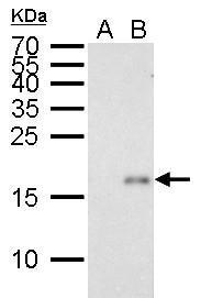
p21 Cip1 antibody [GT8611] detects p21 Cip1 protein by Western blot analysis. A. 30 µg HCT116 whole cell lysate/extract (untreated) B. 30 µg HCT116 whole cell lysate/extract (30 µM Cisplatin treatment for 24 hr) 15 % SDS-PAGE p21 Cip1 antibody [GT8611] dilution: 1:1000Application
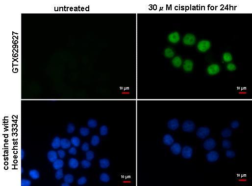
p21 Cip1 antibody [GT8611] detects p21 Cip1 protein at nucleus by immunofluorescent analysis. Sample: HCT116 cells treated with 30μM cisplatin for 24hr and were fixed in 4% paraformaldehyde at RT for 15 min. Green: p21 Cip1 protein stained by p21 Cip1 antibody [GT8611] diluted at 1:500. Blue: Hoechst 33342 staining.Application
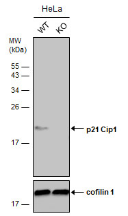
Wild-type (WT) and p21 Cip1 knockout (KO) HeLa cell extracts (30 µg) were separated by 15% SDS-PAGE, and the membrane was blotted with p21 Cip1 antibody [GT8611] diluted at 1:500. The HRP-conjugated anti-mouse IgG antibody was used to detect the primary antibody, and the signal was developed with Trident ECL plus-Enhanced.| Product Name | p21 Cip1 antibody |
|---|---|
| Antibody Type | Primary Antibodies |
| Product description | Mouse Monoclonal antibody [GT8611] to p21 Cip1 (cyclin-dependent kinase inhibitor 1A (p21, Cip1))This gene encodes a potent cyclin-dependent kinase inhibitor. The encoded protein binds to and inhibits the activity of cyclin-CDK2 or -CDK4 complexes, and thus functions as a regulator of cell cycle progression at G1. The expression of this gene is tightly controlled by the tumor suppressor protein p53, through which this protein mediates the p53-dependent cell cycle G1 phase arrest in response to a variety of stress stimuli. This protein can interact with proliferating cell nuclear antigen (PCNA), a DNA polymerase accessory factor, and plays a regulatory role in S phase DNA replication and DNA damage repair. This protein was reported to be specifically cleaved by CASP3-like caspases, which thus leads to a dramatic activation of CDK2, and may be instrumental in the execution of apoptosis following caspase activation. Two alternatively spliced variants, which encode an identical protein, have been reported. [provided by RefSeq] |
| Immunogen | Recombinant protein encompassing a sequence within the center region of human p21 Cip1. The exact sequence is proprietary. |
| Clonality | Monoclonal |
|---|---|
| Isotype | IgG2 |
| Clone Number | GT8611 |
| Host Species | Mouse |
| Tested Applications | ICC/IFWB |
| *Optimal dilutions/concentrations should be determined by the researcher.
: |
|
| Species Reactivity | Human |
| Predicted Reactivity | Cat, Sheep, Rhesus Monkey, BovineCat (84%), Sheep (82%), Rhesus Monkey (98%), Bovine (83%) |
| Concentration | 1mg/ml (Please refer to the vial label for the specific concentration) |
| Purification | Affinity purified |
| Gene Synonyms | wild-type p53-activated fragment 1 antibody p21CIP1 antibody melanoma differentiation associated protein 6 antibody cyclin-dependent kinase inhibitor 1A (p21 Cip1) antibody cyclin-dependent kinase inhibitor 1 antibody WAF1 antibody SDI1 antibo |
|---|---|
| Molecular Weight(MW) | 18 kDa (note) (5) |
| Cellular Localization | Cytoplasm , Nucleus |
Application
p21 Cip1 antibody [GT8611] detects p21 Cip1 protein by Western blot analysis. A. 30 µg HCT116 whole cell lysate/extract (untreated) B. 30 µg HCT116 whole cell lysate/extract (30 µM Cisplatin treatment for 24 hr) 15 % SDS-PAGE p21 Cip1 antibody [GT8611] dilution: 1:1000
Application
p21 Cip1 antibody [GT8611] detects p21 Cip1 protein at nucleus by immunofluorescent analysis. Sample: HCT116 cells treated with 30μM cisplatin for 24hr and were fixed in 4% paraformaldehyde at RT for 15 min. Green: p21 Cip1 protein stained by p21 Cip1 antibody [GT8611] diluted at 1:500. Blue: Hoechst 33342 staining.
Application
Wild-type (WT) and p21 Cip1 knockout (KO) HeLa cell extracts (30 µg) were separated by 15% SDS-PAGE, and the membrane was blotted with p21 Cip1 antibody [GT8611] diluted at 1:500. The HRP-conjugated anti-mouse IgG antibody was used to detect the primary antibody, and the signal was developed with Trident ECL plus-Enhanced.| Positive Control | HCT116 (30 µM Cisplatin treatment for 24 hr) |
|---|---|
| Application Notes | *Optimal dilutions/concentrations should be determined by the researcher.
: |
| Form | Liquid |
|---|---|
| Storage Instructions | Keep as concentrated solution. Aliquot and store at -20ºC or below. Avoid multiple freeze-thaw cycles. |
| Storage Buffer | PBS |


