Application
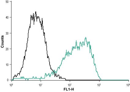
Anti-P2X1_Receptor_(extracellular) - Indirect flow cytometry analysis of MEG-O1 cells: ___ Unstained cells.___ Cells + Anti-P2X1 Receptor (extracellular) antibody, (10 μg antibody/1x106 cells).Application
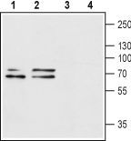
Anti-P2X1_Receptor_(extracellular) - Western blot analysis of rat brain (lanes 1 and 3) and mouse brain (lanes 2 and 4) lysates: 1, 2. Anti-P2X1 Receptor (extracellular) antibody, (1:200). 3, 4. Anti-P2X1 Receptor (extracellular) antibody, preincubated with the control peptide antigen.Application
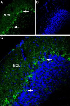
Anti-P2X1_Receptor_(extracellular) - Expression of P2X1 in mouse cerebellum Immunohistochemical staining of mouse cerebellum using Anti-P2X1 Receptor (extracellular) antibody. A. Most of P2X1 labeling (green) appears in fine processes in the molecular layer (MOL) and in Purkinje cells (arrows show examples). B. DAPI is used as the counterstain (blue). C. Merge of A and B.Application
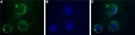
Anti-P2X1_Receptor_(extracellular) - Expression of P2X1 in human SH-SYS5 cells Immunocytochemical staining of intact living human SH-SYS5 cells. A. Extracellular staining of cells with Anti-P2X1 Receptor (extracellular) antibody, (1:50) followed by goat anti-rabbit-AlexaFluor-488 secondary antibody. B. Nuclear staining DAPI as the counterstain. C. Merged images of A and B.| Product Name | P2X1 antibody |
|---|---|
| Antibody Type | Primary Antibodies |
| Product description | Rabbit Polyclonal antibody to P2X1The product of this gene belongs to the family of purinoceptors for ATP. This receptor functions as a ligand-gated ion channel with relatively high calcium permeability. Binding to ATP mediates synaptic transmission between neurons and from neurons to smooth muscle, being responsible, for example, for sympathetic vasoconstriction in small arteries, arterioles and vas deferens. Mouse studies suggest that this receptor is essential for normal male reproductive function. It is possible that the development of selective antagonists for this receptor may provide an effective non-hormonal male contraceptive pill. [provided by RefSeq, Jul 2008] |
| Immunogen | Peptide CRPIYEFHGLYEEK, corresponding to amino acid residues 270-283 of human P2X1 receptor (Accession P51575 ). Extracellular loop. |
| Clonality | Polyclonal |
|---|---|
| Isotype | IgG |
| Host Species | Rabbit |
| Tested Applications | FACSICC/IFIHCWB |
| FACS:1:200~1:500 ICC/IF:1:200~1:500 IHC:1:50~1:200 WB:1:500~1:2000 Notes:Optimal dilutions/concentrations should be determined by the researcher. |
|
| Species Reactivity | HumanMouseRat |
| Concentration | 1mg/ml |
| Purification | Affinity purified |
| Gene Synonyms | purinergic receptor P2X ligand-gated ion channel 1 P2RX1 |
|---|---|
| Target | P2X1 |
Application
Anti-P2X1_Receptor_(extracellular) - Indirect flow cytometry analysis of MEG-O1 cells: ___ Unstained cells.___ Cells + Anti-P2X1 Receptor (extracellular) antibody, (10 μg antibody/1x106 cells).
Application
Anti-P2X1_Receptor_(extracellular) - Western blot analysis of rat brain (lanes 1 and 3) and mouse brain (lanes 2 and 4) lysates: 1, 2. Anti-P2X1 Receptor (extracellular) antibody, (1:200). 3, 4. Anti-P2X1 Receptor (extracellular) antibody, preincubated with the control peptide antigen.
Application
Anti-P2X1_Receptor_(extracellular) - Expression of P2X1 in mouse cerebellum Immunohistochemical staining of mouse cerebellum using Anti-P2X1 Receptor (extracellular) antibody. A. Most of P2X1 labeling (green) appears in fine processes in the molecular layer (MOL) and in Purkinje cells (arrows show examples). B. DAPI is used as the counterstain (blue). C. Merge of A and B.
Application
Anti-P2X1_Receptor_(extracellular) - Expression of P2X1 in human SH-SYS5 cells Immunocytochemical staining of intact living human SH-SYS5 cells. A. Extracellular staining of cells with Anti-P2X1 Receptor (extracellular) antibody, (1:50) followed by goat anti-rabbit-AlexaFluor-488 secondary antibody. B. Nuclear staining DAPI as the counterstain. C. Merged images of A and B.| Application Notes | FACS:1:200~1:500 ICC/IF:1:200~1:500 IHC:1:50~1:200 WB:1:500~1:2000 Notes:Optimal dilutions/concentrations should be determined by the researcher. |
|---|
| Form | Liquid |
|---|---|
| Storage Instructions | Keep as concentrated solution. Aliquot and store at -20ºC or below. Avoid multiple freeze-thaw cycles. |
| Storage Buffer | Phosphate buffered saline (PBS), pH 7.4, 1% BSA, 0.05% NaN3. |


