Application
ApplicationApplication
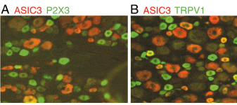
DRG sections double-labeled for ASIC3 (red) and either P2X3 (green, A) or TRPV1 (green, B); double-labeled cells are yellow or orange P2X3 Antibody| Product Name | P2X3 antibody |
|---|---|
| Antibody Type | Primary Antibodies |
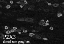
| Product description | Rabbit polyclonal to P2X3 antiserumP2X3 is a subunit in the family of ion channels activated by extracellular ATP. It was cloned from rat dorsal root ganglia. P2X3 has two putative transmembrane domains with intracellular N- and C-termini. It can form homomeric channels and heteromeric channels with other members of the family. P2X3 is expressed in nociceptive sensory neurons. |
| Immunogen | VEKQSTDSGAYSIG Corresponding to residues 383-397 of the carboxy-terminus of rat P2X3 |
| Clonality | Polyclonal |
|---|---|
| Host Species | Rabbit |
| Tested Applications | ICC/IFIHCWB |
| Immunohistochemistry 1:1000 Immunocytochemistry 1:1000 Western Blotting 1:1000 Dilutions listed only as a recommendation. Optimal dilution should be determined by investigator. Immunohistochemistry: Antiserum was used on perfusion fixed tissue. Perfusion: 1) calcium-free Tyrode’s solution, 2) paraformaldehyde-picric acid fixative, and 3) 10% sucrose in PBS as a cryo-protectant. Desired tissues were dissected and stored overnight in 10% sucrose in PBS. Slide-mounted tissue sections were processed for indirect immunofluorescence. Slides were incubated with blocking buffer for 1 hour at room temperature. Primary antiserum was diluted with blocking buffer to the appropriate working concentration. Blocking buffer was removed and slides were incubated for 18-24 hours at 4oC with primary antiserum. Slides were rinsed 3 times and then incubated with secondary antibodies for 1 hour at room temperature. Slides were again rinsed 3 times and coverslipped. Staining was examined using fluorescence microscopy. Immunocytochemistry: P2X3 transfected cells were processed for indirect immunofluorescence. Media was removed and cells were gently washed 3 times with serum-free media. Following fixation procedure, cells were processed for indirect immunofluorescence as described above. Western Blotting: Cell membrane extracts were examined by electrophoresis (8% acrylamide) with SDS under reducing conditions and transferred to a nylon membrane. Membranes were blocked for 1 hour at 4?C with 0.1% Tween 20 and 2.5% milk powder (w/v) in PBS. Membranes were incubated with primary antiserum (1:500 in the same buffer overnight at 4?C. Membranes were rinsed and incubated with horseradish peroxidase conjugated secondary antibody for 1 hour at room temperature. Following rinsing, the membranes were processed using enhanced chemiluminescence.: |
|
| Species Reactivity | HumanRatPrimate |
| Concentration | 1mg/ml |
| Target | P2X3 |
|---|
Application
Application
Application
DRG sections double-labeled for ASIC3 (red) and either P2X3 (green, A) or TRPV1 (green, B); double-labeled cells are yellow or orange P2X3 Antibody| Application Notes | Immunohistochemistry 1:1000 Immunocytochemistry 1:1000 Western Blotting 1:1000 Dilutions listed only as a recommendation. Optimal dilution should be determined by investigator. Immunohistochemistry: Antiserum was used on perfusion fixed tissue. Perfusion: 1) calcium-free Tyrode’s solution, 2) paraformaldehyde-picric acid fixative, and 3) 10% sucrose in PBS as a cryo-protectant. Desired tissues were dissected and stored overnight in 10% sucrose in PBS. Slide-mounted tissue sections were processed for indirect immunofluorescence. Slides were incubated with blocking buffer for 1 hour at room temperature. Primary antiserum was diluted with blocking buffer to the appropriate working concentration. Blocking buffer was removed and slides were incubated for 18-24 hours at 4oC with primary antiserum. Slides were rinsed 3 times and then incubated with secondary antibodies for 1 hour at room temperature. Slides were again rinsed 3 times and coverslipped. Staining was examined using fluorescence microscopy. Immunocytochemistry: P2X3 transfected cells were processed for indirect immunofluorescence. Media was removed and cells were gently washed 3 times with serum-free media. Following fixation procedure, cells were processed for indirect immunofluorescence as described above. Western Blotting: Cell membrane extracts were examined by electrophoresis (8% acrylamide) with SDS under reducing conditions and transferred to a nylon membrane. Membranes were blocked for 1 hour at 4?C with 0.1% Tween 20 and 2.5% milk powder (w/v) in PBS. Membranes were incubated with primary antiserum (1:500 in the same buffer overnight at 4?C. Membranes were rinsed and incubated with horseradish peroxidase conjugated secondary antibody for 1 hour at room temperature. Following rinsing, the membranes were processed using enhanced chemiluminescence.: |
|---|
| Form | Liquid |
|---|---|
| Storage Instructions | Upon Receipt - Keep as concentrated solution. Aliquot and store at -20ºC or below. Avoid freeze-thaw cycles. |
| Storage Buffer | Whole Serum |


