Application
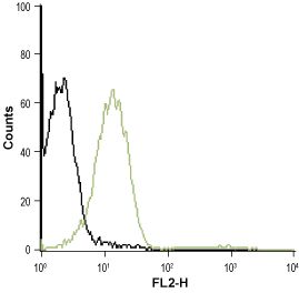
Anti-P2Y1_Receptor_(extracellular) - Indirect flow cytometry analysis of Jurkat (human acute T cell leukemia) living cells: ___ Unstained cells.___ Cells stained + Anti-P2Y1 Receptor (extracellular) antibody, (5 μg/5x105 cells).Application
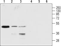
Anti-P2Y1_Receptor_(extracellular) - Western blot analysis of rat brain (lanes 1 and 4), Jurkat (lanes 2 and 5) and K-562 (lanes 3 and 6) lysates: 1-3. Anti-P2Y1 Receptor (extracellular) antibody, (1:200). 4-6. Anti-P2Y1 Receptor (extracellular) antibody, preincubated with the control peptide antigen.Application
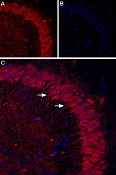
Anti-P2Y1_Receptor_(extracellular) - Expression of P2Y1 in mouse hippocampus Immunohistochemical staining of mouse hippocampal CA3 region using Anti-P2Y1 (extracellular) antibody. A. P2Y1 staining (red) appears in the pyramidal layer (arrows). B. Nuclei staining using DAPI as the counterstain (blue). C. Merged images of panels A and B.Application
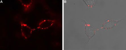
Anti-P2Y1_Receptor_(extracellular) - Expression of P2Y1 receptor in rat PC12 cells Immunocytochemical staining of intact living rat pheochromocytoma PC12 cells. A. Extracellular staining of cells using Anti-P2Y1 Receptor (extracellular) antibody, (1:50) followed by goat anti-rabbit-AlexaFluor-594 secondary antibody (red). B. Merge of extracellular staining with live view of the cells.| Product Name | P2Y1 antibody |
|---|---|
| Antibody Type | Primary Antibodies |
| Product description | Rabbit Polyclonal antibody to P2Y1The product of this gene belongs to the family of G-protein coupled receptors. This family has several receptor subtypes with different pharmacological selectivity, which overlaps in some cases, for various adenosine and uridine nucleotides. This receptor functions as a receptor for extracellular ATP and ADP. In platelets binding to ADP leads to mobilization of intracellular calcium ions via activation of phospholipase C, a change in platelet shape, and probably to platelet aggregation. [provided by RefSeq, Jul 2008] |
| Immunogen | Peptide SDEYLRSYFIYSMC, corresponding to amino acid residues 207-220 of human P2Y1 Receptor (Accession P47900 ). 2nd extracellular loop. |
| Clonality | Polyclonal |
|---|---|
| Isotype | IgG |
| Host Species | Rabbit |
| Tested Applications | FACSICC/IFIHCWB |
| FACS:1:200~1:500 ICC/IF:1:200~1:500 IHC:1:50~1:200 WB:1:500~1:2000 Notes:Optimal dilutions/concentrations should be determined by the researcher. |
|
| Species Reactivity | HumanMouseRat |
| Concentration | 1mg/ml |
| Purification | Affinity purified |
| Gene Synonyms | purinergic receptor P2Y G-protein coupled 1 P2RY1 |
|---|---|
| Target | P2Y1 |
Application
Anti-P2Y1_Receptor_(extracellular) - Indirect flow cytometry analysis of Jurkat (human acute T cell leukemia) living cells: ___ Unstained cells.___ Cells stained + Anti-P2Y1 Receptor (extracellular) antibody, (5 μg/5x105 cells).
Application
Anti-P2Y1_Receptor_(extracellular) - Western blot analysis of rat brain (lanes 1 and 4), Jurkat (lanes 2 and 5) and K-562 (lanes 3 and 6) lysates: 1-3. Anti-P2Y1 Receptor (extracellular) antibody, (1:200). 4-6. Anti-P2Y1 Receptor (extracellular) antibody, preincubated with the control peptide antigen.
Application
Anti-P2Y1_Receptor_(extracellular) - Expression of P2Y1 in mouse hippocampus Immunohistochemical staining of mouse hippocampal CA3 region using Anti-P2Y1 (extracellular) antibody. A. P2Y1 staining (red) appears in the pyramidal layer (arrows). B. Nuclei staining using DAPI as the counterstain (blue). C. Merged images of panels A and B.
Application
Anti-P2Y1_Receptor_(extracellular) - Expression of P2Y1 receptor in rat PC12 cells Immunocytochemical staining of intact living rat pheochromocytoma PC12 cells. A. Extracellular staining of cells using Anti-P2Y1 Receptor (extracellular) antibody, (1:50) followed by goat anti-rabbit-AlexaFluor-594 secondary antibody (red). B. Merge of extracellular staining with live view of the cells.| Application Notes | FACS:1:200~1:500 ICC/IF:1:200~1:500 IHC:1:50~1:200 WB:1:500~1:2000 Notes:Optimal dilutions/concentrations should be determined by the researcher. |
|---|
| Form | Liquid |
|---|---|
| Storage Instructions | Keep as concentrated solution. Aliquot and store at -20ºC or below. Avoid multiple freeze-thaw cycles. |
| Storage Buffer | Phosphate buffered saline (PBS), pH 7.4, 1% BSA, 0.05% NaN3. |


