Application
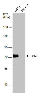
Various whole cell extracts (30 µg) were separated by 7.5% SDS-PAGE, and the membrane was blotted with p63 antibody [N2C1], Internal diluted at 1:1000. The HRP-conjugated anti-rabbit IgG antibody was used to detect the primary antibody.Application
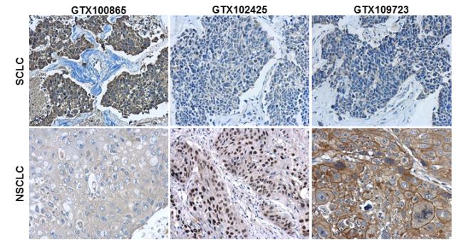
Immunohistochemical characterization of Synaptophysin , p63 and Cytokeratin 7 in human small cell lung cancer (SCLC) and non-small cell lung cancer (NSCLC) specimens. Sample: Paraffin-embedded human SCLC (upper panel) and NSCLC (lower panel). The section was pre-treated using heat mediated antigen retrieval with sodium citrate buffer (pH6) for 15 mins. The section was then incubated with primary antibody at 1:500 overnight at 4℃ and detected using an HRP conjugated avidin-biotin-peroxidase Complex system. DAB was used as the chromogen and counterstained with haematoxylin.Application
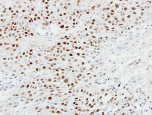
Immunohistochemical analysis of paraffin-embedded SCC4 xenograft, using p63 antibody at 1:100 dilution.Application
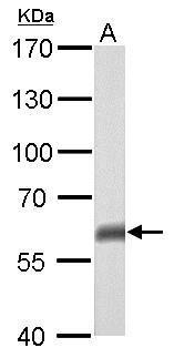
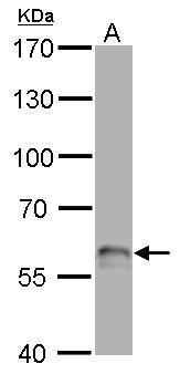
p63 antibody [N2C1], Internal detects TP63 protein by western blot analysis. A. 50 µg mouse brain lysate/extract 7.5% SDS-PAGE p63 antibody [N2C1], Internal dilution: 1:500 The HRP-conjugated anti-rabbit IgG antibody was used to detect the primary antibody.Application
p63 antibody [N2C1], Internal detects TP63 protein by western blot analysis. A. 50 µg rat brain lysate/extract 7.5% SDS-PAGE p63 antibody [N2C1], Internal dilution: 1:500 The HRP-conjugated anti-rabbit IgG antibody was used to detect the primary antibody.Application
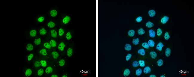
p63 antibody [N2C1], Internal detects p63 protein at nucleus by immunofluorescent analysis. Sample: A431 cells were fixed in 4% paraformaldehyde at RT for 15 min. Green: p63 protein stained by p63 antibody [N2C1], Internal diluted at 1:500. Blue: Hoechst 33342 staining. Scale bar = 10 μm.Application
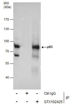
Immunoprecipitation of p63 protein from A431 whole cell extracts using 5 μg of p63 antibody [N2C1], Internal . Western blot analysis was performed using p63 antibody [N2C1], Internal . EasyBlot anti-Rabbit IgG was used as a secondary reagent.| Product Name | p63 antibody , Internal |
|---|---|
| Antibody Type | Primary Antibodies |
| Product description | Rabbit Polyclonal antibody to p63 (tumor protein p63)This gene encodes a member of the p53 family of transcription factors. An animal model, p63 -/- mice, has been useful in defining the role this protein plays in the development and maintenance of stratified epithelial tissues. p63 -/- mice have several developmental defects which include the lack of limbs and other tissues, such as teeth and mammary glands, which develop as a result of interactions between mesenchyme and epithelium. Mutations in this gene are associated with ectodermal dysplasia, and cleft lip/palate syndrome 3 (EEC3); split-hand/foot malformation 4 (SHFM4); ankyloblepharon-ectodermal defects-cleft lip/palate; ADULT syndrome (acro-dermato-ungual-lacrimal-tooth); limb-mammary syndrome; Rap-Hodgkin syndrome (RHS); and orofacial cleft 8. Both alternative splicing and the use of alternative promoters results in multiple transcript variants encoding different proteins. Many transcripts encoding different proteins have been reported but the biological validity and the full-length nature of these variants have not been determined. [provided by RefSeq] |
| Immunogen | Recombinant protein encompassing a sequence within the center region of human p63. The exact sequence is proprietary. |
| Clonality | Polyclonal |
|---|---|
| Isotype | IgG |
| Host Species | Rabbit |
| Tested Applications | ICC/IFIHC-PIPWB |
| *Optimal dilutions/concentrations should be determined by the researcher.
: |
|
| Species Reactivity | HumanMouseRat |
| Predicted Reactivity | Zebrafish, Xenopus laevis, Chicken, BovineZebrafish (91%), Xenopus laevis (94%), Chicken (96%), Bovine (100%) |
| Concentration | 1mg/ml (Please refer to the vial label for the specific concentration) |
| Purification | Affinity purified |
| Gene Synonyms | AIS antibody B(p51A) antibody B(p51B) antibody EEC3 antibody KET antibody LMS antibody NBP antibody OFC8 antibody RHS antibody SHFM4 antibody TP53CP antibody TP53L antibody TP73L antibody p40 antibody p51 antibody p53CP antibody p63 antibod |
|---|---|
| Molecular Weight(MW) | 77 kDa (note) (11) |
| Cellular Localization | Nucleus |
Application
Various whole cell extracts (30 µg) were separated by 7.5% SDS-PAGE, and the membrane was blotted with p63 antibody [N2C1], Internal diluted at 1:1000. The HRP-conjugated anti-rabbit IgG antibody was used to detect the primary antibody.
Application
Immunohistochemical characterization of Synaptophysin , p63 and Cytokeratin 7 in human small cell lung cancer (SCLC) and non-small cell lung cancer (NSCLC) specimens. Sample: Paraffin-embedded human SCLC (upper panel) and NSCLC (lower panel). The section was pre-treated using heat mediated antigen retrieval with sodium citrate buffer (pH6) for 15 mins. The section was then incubated with primary antibody at 1:500 overnight at 4℃ and detected using an HRP conjugated avidin-biotin-peroxidase Complex system. DAB was used as the chromogen and counterstained with haematoxylin.
Application
Immunohistochemical analysis of paraffin-embedded SCC4 xenograft, using p63 antibody at 1:100 dilution.
Application
p63 antibody [N2C1], Internal detects TP63 protein by western blot analysis. A. 50 µg mouse brain lysate/extract 7.5% SDS-PAGE p63 antibody [N2C1], Internal dilution: 1:500 The HRP-conjugated anti-rabbit IgG antibody was used to detect the primary antibody.
Application
p63 antibody [N2C1], Internal detects TP63 protein by western blot analysis. A. 50 µg rat brain lysate/extract 7.5% SDS-PAGE p63 antibody [N2C1], Internal dilution: 1:500 The HRP-conjugated anti-rabbit IgG antibody was used to detect the primary antibody.
Application
p63 antibody [N2C1], Internal detects p63 protein at nucleus by immunofluorescent analysis. Sample: A431 cells were fixed in 4% paraformaldehyde at RT for 15 min. Green: p63 protein stained by p63 antibody [N2C1], Internal diluted at 1:500. Blue: Hoechst 33342 staining. Scale bar = 10 μm.
Application
Immunoprecipitation of p63 protein from A431 whole cell extracts using 5 μg of p63 antibody [N2C1], Internal . Western blot analysis was performed using p63 antibody [N2C1], Internal . EasyBlot anti-Rabbit IgG was used as a secondary reagent.| Positive Control | A431 , mouse brain , rat brain |
|---|---|
| Application Notes | *Optimal dilutions/concentrations should be determined by the researcher.
: |
| Form | Liquid |
|---|---|
| Storage Instructions | Keep as concentrated solution. Aliquot and store at -20ºC or below. Avoid multiple freeze-thaw cycles. |
| Storage Buffer | 1XPBS, 20% Glycerol (pH7). 0.01% Thimerosal was added as a preservative. |


