Application
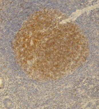
Immunohistochemistry staining (paraffin sections) of PAG/Cbp in germinal center of lymphatic follicle and in dispersed T cells in appendix tissue by monoclonal antibody Positive signal in T cells.Application
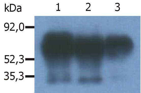
Immunoprecipitation of human PAG/Cbp from the lysate of RAJI human Burkitt lymphoma cell line. Western blot was immunostained with anti-human PAG . Note: PAG/Cbp is a 46 kDa adaptor protein, which however migrates on SDS PAGE gels anomalously as an 80 kDa molecule. Lane 1,2: immunoprecipitation with anti-PAG Lane 3: immunoprecipitation with anti-PAG| Product Name | PAG antibody |
|---|---|
| Antibody Type | Primary Antibodies |
| Product description | Mouse monoclonal [PAG - C1] to PAGCsk binding protein, also known as PAG (phosphoprotein associated with GEMs), is a ubiquitously expressed 46 kDa transmembrane adaptor protein present in membrane rafts (glycosphingolipid-enriched microdomains), which however migrates on SDS PAGE gels anomalously as an 80 kDa molecule. Following tyrosine phosphorylation by Src family kinases, PAG binds and thereby activates the protein tyrosine kinase Csk, the major negative regulator of the Src family kinases. Signaling via the B-cell receptor in B cells or high affinity IgE receptor (FcepsilonRI) in mast cells leads to PAG increased tyrosine phosphorylation and Csk binding, while T cell receptor signaling causes PAG dephosphorylation, loss of Csk binding and increased activation of the protein tyrosine kinase Lck. |
| Immunogen | Synthetic C-teminal peptide (last 15 amino acids) (Human) conjugated to KLH. |
| Clonality | Monoclonal |
|---|---|
| Isotype | IgG2 |
| Clone Number | PAG-C1 |
| Host Species | Mouse |
| Tested Applications | IHC-PIPWB |
| IP: Use at an assay dependant concentration. Optimal dilutions/concentrations should be determined by the end user.: |
|
| Species Reactivity | HumanMouseRatBovine |
| Concentration | 1mg/ml (Please refer to the vial label for the specific concentration) |
| Purification | Affinity purified |
| Gene Synonyms | CBP FLJ37858 MGC138364 PAG |
|---|---|
| Target | PAG |
| Tissue Specificity | The antibody recognizes an epitope located in the C-terminal domain of Csk-binding protein (Cbp), also known as protein associated with glycosphingolipid-enriched microdomains (PAG). |
Application
Immunohistochemistry staining (paraffin sections) of PAG/Cbp in germinal center of lymphatic follicle and in dispersed T cells in appendix tissue by monoclonal antibody Positive signal in T cells.
Application
Immunoprecipitation of human PAG/Cbp from the lysate of RAJI human Burkitt lymphoma cell line. Western blot was immunostained with anti-human PAG . Note: PAG/Cbp is a 46 kDa adaptor protein, which however migrates on SDS PAGE gels anomalously as an 80 kDa molecule. Lane 1,2: immunoprecipitation with anti-PAG Lane 3: immunoprecipitation with anti-PAG| Application Notes | IP: Use at an assay dependant concentration. Optimal dilutions/concentrations should be determined by the end user.: |
|---|
| Form | Liquid |
|---|---|
| Storage Instructions | Keep as concentrated solution. Store at 4ºC short term. For extended storage aliquot and store at -20ºC or below. Avoid freeze-thaw cycles. |
| Storage Buffer | Phosphate buffered saline pH 7.4 containing 15mM sodium azide |


