Application
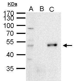
PAX6 antibody immunoprecipitates PAX6 protein in IP experiments. IP Sample: 1000 µg 293T whole cell lysate/extract A. 50 μg 293T whole cell lysate/extract B. Control with 2 μg of preimmune rabbit IgG C. Immunoprecipitation of PAX6 protein by 2 μg of PAX6 antibody 10% SDS-PAGE The immunoprecipitated PAX6 protein was detected by PAX6 antibody diluted at 1:1000. EasyBlot anti-rabbit IgG was used as a secondary reagent.Application
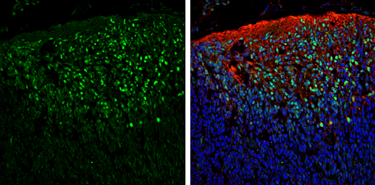
PAX6 antibody detects PAX6 protein expression by immunohistochemical analysis. Sample: Frozen sectioned E13.5 Rat brain. Green: PAX6 protein stained by PAX6 antibody diluted at 1:250. Red: beta Tubulin 3/ TUJ1, a mature neuron marker, stained by beta Tubulin 3/ TUJ1 antibody [GT11710] diluted at 1:500. Blue: Fluoroshield with DAPI .Application
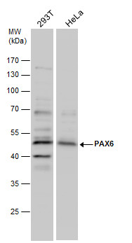
PAX6 antibody detects PAX6 protein by Western blot analysis. Various whole cell extracts (30 μg) were separated by 10% SDS-PAGE, and the membrane was blotted with PAX6 antibody diluted by 1:1000.Application
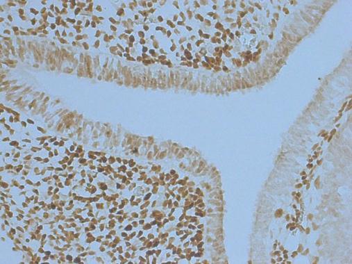
PAX6 antibody detects PAX6 protein at nucleus in embryonic mouse brain by immunohistochemical analysis. Sample: Frozen section of embryonic mouse brain (mE12.5). PAX6 antibody diluted at 1:250.Application
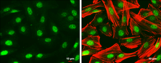
PAX6 antibody detects PAX6 protein at nucleus by immunofluorescent analysis. Sample: SK-N-SH cells were fixed in 4% paraformaldehyde at RT for 15 min. Green: PAX6 protein stained by PAX6 antibody diluted at 1:500. Red: Phalloidin, a cytoskeleton marker, diluted at 1:50. Scale bar = 10 μm.Application
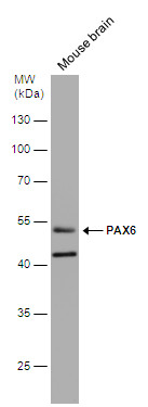
Mouse tissue extract (50 µg) was separated by 10% SDS-PAGE, and the membrane was blotted with PAX6 antibody diluted at 1:1000. The HRP-conjugated anti-rabbit IgG antibody was used to detect the primary antibody.Application
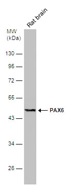
Rat tissue extract (50 µg) was separated by 10% SDS-PAGE, and the membrane was blotted with PAX6 antibody diluted at 1:500. The HRP-conjugated anti-rabbit IgG antibody was used to detect the primary antibody.| Product Name | PAX6 antibody |
|---|---|
| Antibody Type | Primary Antibodies |
| Product description | Rabbit Polyclonal antibody to PAX6 (paired box 6)This gene encodes paired box gene 6, one of many human homologs of the Drosophila melanogaster gene prd. In addition to the hallmark feature of this gene family, a conserved paired box domain, the encoded protein also contains a homeo box domain. Both domains are known to bind DNA, and function as regulators of gene transcription. This gene is expressed in the developing nervous system, and in developing eyes. Mutations in this gene are known to cause ocular disorders such as aniridia and Peter's anomaly. Alternatively spliced transcript variants encoding either the same or different isoform have been found for this gene. [provided by RefSeq] |
| Immunogen | Recombinant protein encompassing a sequence within the center region of human PAX6. The exact sequence is proprietary. |
| Clonality | Polyclonal |
|---|---|
| Isotype | IgG |
| Host Species | Rabbit |
| Tested Applications | ICC/IFIHC-FIPWB |
| *Optimal dilutions/concentrations should be determined by the researcher.
: |
|
| Species Reactivity | HumanMouseRat |
| Predicted Reactivity | Xenopus laevis, Dog, Pig, Rabbit, Chicken, Rhesus Monkey, BovineXenopus laevis (94%), Dog (100%), Pig (100%), Rabbit (99%), Chicken (98%), Rhesus Monkey (100%), Bovine (100%) |
| Concentration | 1mg/ml (Please refer to the vial label for the specific concentration) |
| Purification | Affinity purified |
| Gene Synonyms | AN antibody AN2 antibody D11S812E antibody MGC17209 antibody MGDA antibody WAGR antibody PAX6 antibody paired box protein Pax-6 antibody aniridia type II protein antibody oculorhombin antibody paired box homeotic gene-6 antibody paired box 6 ant |
|---|---|
| Molecular Weight(MW) | 47 kDa (note) (10) |
| Cellular Localization | Nucleus |
Application
PAX6 antibody immunoprecipitates PAX6 protein in IP experiments. IP Sample: 1000 µg 293T whole cell lysate/extract A. 50 μg 293T whole cell lysate/extract B. Control with 2 μg of preimmune rabbit IgG C. Immunoprecipitation of PAX6 protein by 2 μg of PAX6 antibody 10% SDS-PAGE The immunoprecipitated PAX6 protein was detected by PAX6 antibody diluted at 1:1000. EasyBlot anti-rabbit IgG was used as a secondary reagent.
Application
PAX6 antibody detects PAX6 protein expression by immunohistochemical analysis. Sample: Frozen sectioned E13.5 Rat brain. Green: PAX6 protein stained by PAX6 antibody diluted at 1:250. Red: beta Tubulin 3/ TUJ1, a mature neuron marker, stained by beta Tubulin 3/ TUJ1 antibody [GT11710] diluted at 1:500. Blue: Fluoroshield with DAPI .
Application
PAX6 antibody detects PAX6 protein by Western blot analysis. Various whole cell extracts (30 μg) were separated by 10% SDS-PAGE, and the membrane was blotted with PAX6 antibody diluted by 1:1000.
Application
PAX6 antibody detects PAX6 protein at nucleus in embryonic mouse brain by immunohistochemical analysis. Sample: Frozen section of embryonic mouse brain (mE12.5). PAX6 antibody diluted at 1:250.
Application
PAX6 antibody detects PAX6 protein at nucleus by immunofluorescent analysis. Sample: SK-N-SH cells were fixed in 4% paraformaldehyde at RT for 15 min. Green: PAX6 protein stained by PAX6 antibody diluted at 1:500. Red: Phalloidin, a cytoskeleton marker, diluted at 1:50. Scale bar = 10 μm.
Application
Mouse tissue extract (50 µg) was separated by 10% SDS-PAGE, and the membrane was blotted with PAX6 antibody diluted at 1:1000. The HRP-conjugated anti-rabbit IgG antibody was used to detect the primary antibody.
Application
Rat tissue extract (50 µg) was separated by 10% SDS-PAGE, and the membrane was blotted with PAX6 antibody diluted at 1:500. The HRP-conjugated anti-rabbit IgG antibody was used to detect the primary antibody.| Positive Control | A431 , HepG2 , mouse brain , rat brain |
|---|---|
| Application Notes | *Optimal dilutions/concentrations should be determined by the researcher.
: |
| Form | Liquid |
|---|---|
| Storage Instructions | Keep as concentrated solution. Aliquot and store at -20ºC or below. Avoid multiple freeze-thaw cycles. |
| Storage Buffer | 0.1M Tris, 0.1M Glycine, 10% Glycerol (pH7). 0.01% Thimerosal was added as a preservative. |


